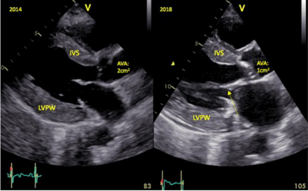
1. This month's #EHJCaseReports #tweetorial focuses on a cardiac condition that can result in these findings 👇 on #echofirst. What could it be?
1 Hypertension
2 Hypertrophic cardiomyopathy
3 Aortic stenosis
4 Cardiac amyloidosis
doi.org/10.1093/ehjcr/…
#CardioTwitter @EHJCREiC
1 Hypertension
2 Hypertrophic cardiomyopathy
3 Aortic stenosis
4 Cardiac amyloidosis
doi.org/10.1093/ehjcr/…
#CardioTwitter @EHJCREiC

2. It’s cardiac amyloidosis! Here is a great resource from @escardio WG on Myocardial and Pericardial Diseases to start with
doi.org/10.1093/eurhea…
doi.org/10.1093/eurhea…
3. First, what is amyloidosis?
Amyloidosis occurs when abnormal proteins misfold and deposit in tissues as beta-pleated sheets. These disrupt tissue structure and can cause damage to many different organs.
Amyloidosis occurs when abnormal proteins misfold and deposit in tissues as beta-pleated sheets. These disrupt tissue structure and can cause damage to many different organs.
4. With regards to cardiac amyloidosis, these abnormal proteins may either be abnormal light chains (AL) or transthyretin (ATTR). Here is a full list of amyloid subtypes that affect the heart. 

5. ATTR may be divided into wild-type or mutant variants. Wild-type refers to the accumulation of the transthyretin protein over time.
Mutant refers to a heritable condition whereby genetic mutations result in destabilisation of TTR.
More about ATTR-CA: doi.org/10.1093/ehjcr/…
Mutant refers to a heritable condition whereby genetic mutations result in destabilisation of TTR.
More about ATTR-CA: doi.org/10.1093/ehjcr/…
6. But how common is cardiac amyloidosis? Recent studies suggest that the prevalence of ATTR-CA is much higher than previously anticipated in older patients with HF.
7. For example, how common do you think ATTR-CA is among patients undergoing TAVR?
8. In this study, ATTR cardiac amyloidosis was present in 16% of patients with severe calcific AS undergoing TAVR! doi.org/10.1093/eurhea… 

9. When should we suspect cardiac amyloidosis?
Common presentations: dyspnoea, oedema or syncope.
doi.org/10.1093/ehjcr/…
Common presentations: dyspnoea, oedema or syncope.
doi.org/10.1093/ehjcr/…

10. Further clues: periorbital purpura, macroglossia, nephrotic syndrome, neuropathy, carpal tunnel syndrome, lumbar spinal stenosis or spontaneous biceps rupture doi.org/10.1093/ehjcr/… 

11. On EKG, they may have low voltage QRS, a ‘pseudo infarct pattern’, atrial arrhythmias, AV nodal conduction block, or a bundle branch block.
doi.org/10.1093/ehjcr/…
doi.org/10.1093/ehjcr/…
12. On echocardiography, there may be increased LV wall thickness without a history of hypertension, RV hypertrophy, ‘speckled’ appearance, thick interatrial septum, pericardial effusion and a bull’s eye appearance on strain imaging.
doi.org/10.1093/ehjcr/…

doi.org/10.1093/ehjcr/…


13. This case report elegantly demonstrates evolution of ECG & echocardiographic changes in cardiac amyloidosis
doi.org/10.1093/ehjcr/…
doi.org/10.1093/ehjcr/…
14. This table provides a summary of cardiac and extracardiac amyloidosis red flags
doi.org/10.1093/eurhea…
doi.org/10.1093/eurhea…

15. How should one investigate suspected cardiac amyloidosis?
Serum and urine immunofixation, as well as a serum free light chain assay, are important to identify AL amyloidosis.
👇
Serum and urine immunofixation, as well as a serum free light chain assay, are important to identify AL amyloidosis.
👇
16. If these labs identify the presence of a monoclonal protein, or if the results are indeterminate, then a biopsy should be performed, which may be from bone marrow, fat or endomyocardium.
17. Why is it so important to identify AL cardiac amyloidosis?
Prognostic markers include troponin T >0.03 mcg/L, NTpro-BNP >1800ng/L, and the difference between involved minus uninvolved serum-free light chains >18mg/dL. 👇
Prognostic markers include troponin T >0.03 mcg/L, NTpro-BNP >1800ng/L, and the difference between involved minus uninvolved serum-free light chains >18mg/dL. 👇
19. Hang on a second though, what happens if those initial labs did NOT identify the presence of a monoclonal protein? Does that mean my patient does not have cardiac amyloid?
20. Unfortunately no.
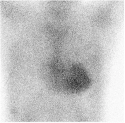
If the initial labs are normal, it is possible the patient may have ATTR cardiac amyloidosis. In this case, a technetium 99 pyrophosphate scan can help clinch the diagnosis.
If the initial labs are normal, it is possible the patient may have ATTR cardiac amyloidosis. In this case, a technetium 99 pyrophosphate scan can help clinch the diagnosis.
21. Using heart to contralateral ratio, a value of ≥1.5 at one hour can be classified as ATTR positive. Furthermore, a semiquantitative visual grading of 2-3 is strongly suggestive of ATTR cardiac amyloidosis. 👇
22. The use of the PYP scan may bypass the need for an endomyocardial biopsy in patients with ATTR cardiac amyloid.
doi.org/10.1093/ehjcr/…
doi.org/10.1093/ehjcr/…

23. This is a nice summary of the diagnostic work-up for patients with suspected cardiac amyloidosis.
doi.org/10.1093/eurhea…
doi.org/10.1093/eurhea…

24. Several trials now demonstrate how targeted treatments focused on different stages of the TTR amyloid cascade can improve outcomes in ATTR patients. 👇
25. Most notable is tafamidis for the treatment of ATTR cardiac amyloid. In the ATTR-ACT trial, tafamidis reduced mortality by 30% compared to placebo with an NNT of 7.5 to prevent one death.
doi.org/10.1093/eurhea…
doi.org/10.1093/eurhea…

Key takeaways from this tweetorial:
▶️Cardiac amyloidosis is not a rare disease
▶️Know when to suspect cardiac amyloidosis
▶️Know what labs to order initially as AL cardiac amyloid must be diagnosed promptly.
▶️Newer therapies for cardiac amyloidosis are effective.
▶️Cardiac amyloidosis is not a rare disease
▶️Know when to suspect cardiac amyloidosis
▶️Know what labs to order initially as AL cardiac amyloid must be diagnosed promptly.
▶️Newer therapies for cardiac amyloidosis are effective.
We hope you enjoyed this tweetorial from the #EHJCaseReports #SoMe team @DrPRao @FarhanaAra @TJ_Yeo @aayshacader @ANazmiCalik 👏
We’ll leave you with an illustrative summary of cardiac amyloidosis doi.org/10.1093/eurhea…
@EHJCREiC @cfcamm #ESCYoung #cardiology
We’ll leave you with an illustrative summary of cardiac amyloidosis doi.org/10.1093/eurhea…
@EHJCREiC @cfcamm #ESCYoung #cardiology

• • •
Missing some Tweet in this thread? You can try to
force a refresh


