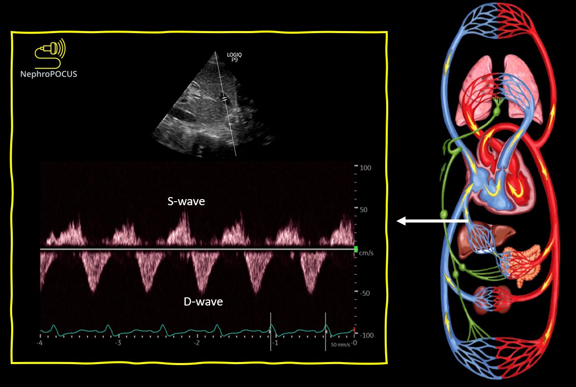
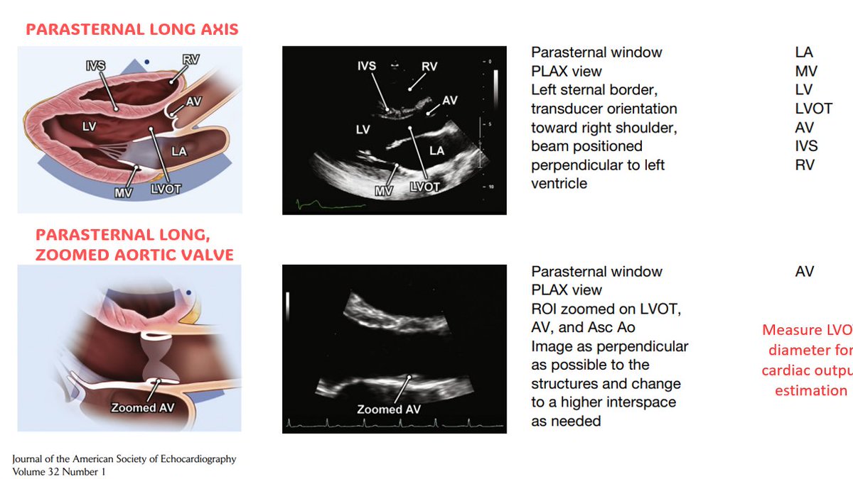
Time to discuss some rationale/evidence behind doing #VExUS #POCUS #Nephrology
A short #tweetorial #MedEd 👇
1/ Is fluid overload harmful?
of course yes. Here is a recent meta-analysis.
A short #tweetorial #MedEd 👇
1/ Is fluid overload harmful?
of course yes. Here is a recent meta-analysis.

2/ Does fluid administration affect renal venous flow in asymptomatic but vulnerable patients (#heartfailure)?
#POCUS #VExUS
#POCUS #VExUS

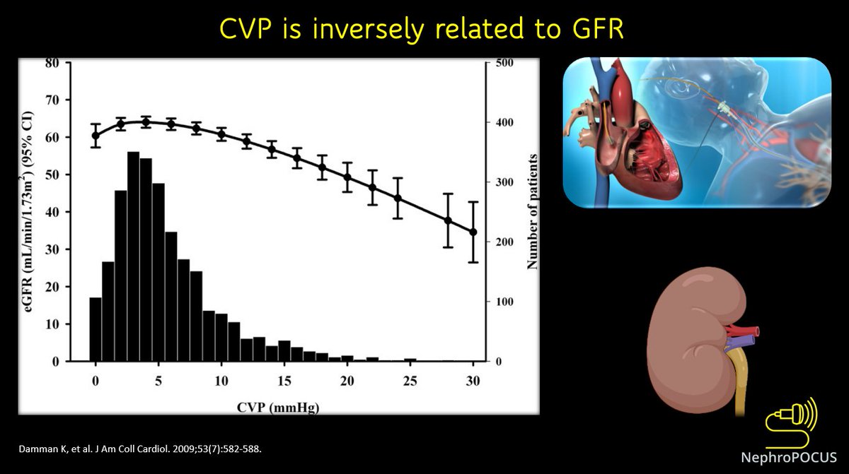
3/ In fact, elevated CVP is associated with reduced GFR.
This 👇is a study in outpatients undergoing right heart cath (N = 2557). In CVP values >6 mm Hg, a steep decrease in GFR was observed.
This 👇is a study in outpatients undergoing right heart cath (N = 2557). In CVP values >6 mm Hg, a steep decrease in GFR was observed.
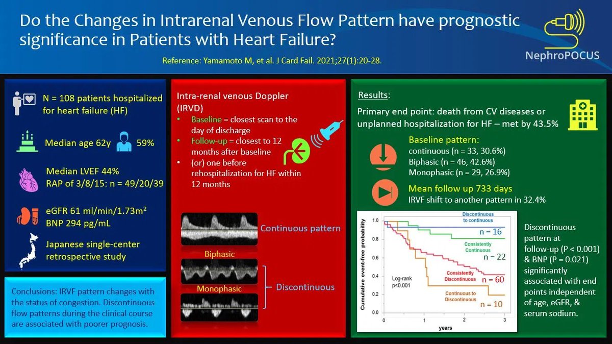
4/ Do alterations in intra-renal venous flow bear prognostic significance in heart failure patients? Yes.
#VExUS
#VExUS

5/ More recent study looking at the effect of change in renal waveform pattern on prognosis.
#VExUS #POCUS
#VExUS #POCUS
6/ Can intra-renal Doppler predict prognosis in patients with chronic pulmonary hypertension? Yes.
Here is a study 👇looking at renal venous stasis index (probably this index is better in chronic settings than observing the pattern)
#VExUS #POCUS
Here is a study 👇looking at renal venous stasis index (probably this index is better in chronic settings than observing the pattern)
#VExUS #POCUS

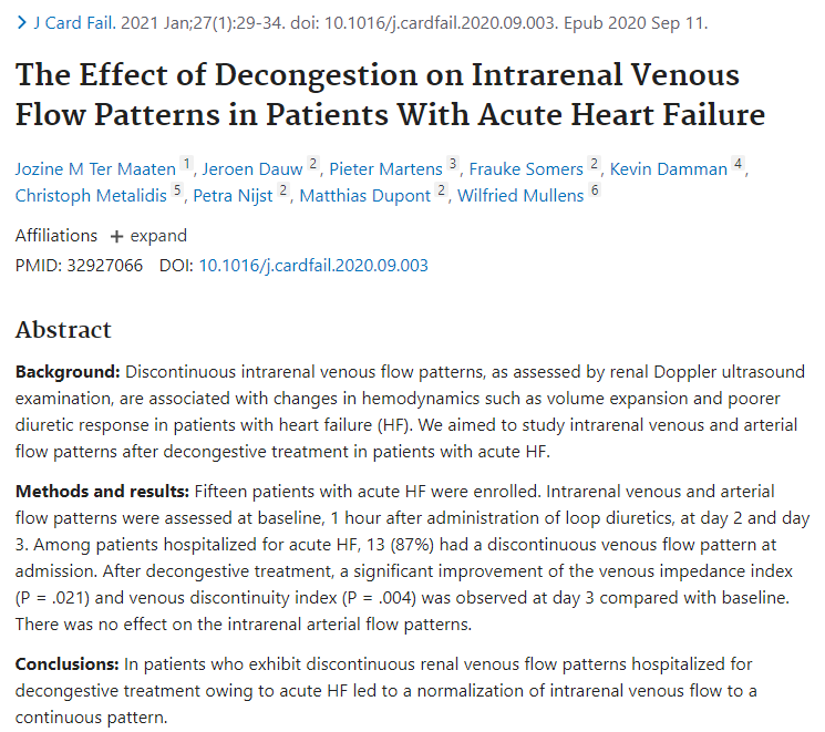
7/ Does decongestive therapy normalize the intra-renal venous flow pattern in patients hospitalized for acute heart failure? Yes.
#POCUS #VExUS
onlinejcf.com/article/S1071-…
#POCUS #VExUS
onlinejcf.com/article/S1071-…
8/ How about portal vein? It is easier to obtain than intra-renal #VExUS
Does it predict AKI? Yes (in cardiac surgery patients 👇)
This is actually the parent study of the VExUS paper (multi-vein congestion assessment)
Does it predict AKI? Yes (in cardiac surgery patients 👇)
This is actually the parent study of the VExUS paper (multi-vein congestion assessment)

9/ Does the portal vein flow change dynamically during Decongestion in Patients with #heartfailure and Cardio-Renal Syndrome? Yes.
Here is a recent case series by our own #VExUS ologist @ArgaizR
karger.com/Article/FullTe…
Here is a recent case series by our own #VExUS ologist @ArgaizR
karger.com/Article/FullTe…

10/ Another small study in critically ill cardiac ICU patients showing improvement in hepatic/portal vein patterns with decongestive therapy.
#POCUS #VExUS
#POCUS #VExUS

11/ Another study that looked at the hepatic/portal/intra-renal #VExUS in predicting adverse renal events. But this is a general ICU cohort (= patients can have AKI due to other causes/multiple factors; not just congestion) 

12/ Finally, the famous #VExUS scoring by @WBeaubien @ThinkingCC @khaycock2 and colleagues.
using IVC #POCUS + hepatic + portal + intra-renal Doppler to quantify venous congestion predicts AKI.
using IVC #POCUS + hepatic + portal + intra-renal Doppler to quantify venous congestion predicts AKI.

13/ Using multiple points overcomes the inherent limitations of individual components as well as technical issues.
Combined #VExUS score predicts AKI better than IVC #POCUS (CVP) alone
Combined #VExUS score predicts AKI better than IVC #POCUS (CVP) alone

14/ Future directions:
Femoral/splenic veins may be used as an adjunct in the assessment of venous congestion.
#VExUS #POCUS
ncbi.nlm.nih.gov/pmc/articles/P…
Femoral/splenic veins may be used as an adjunct in the assessment of venous congestion.
#VExUS #POCUS
ncbi.nlm.nih.gov/pmc/articles/P…
16/ Future directions:
A multi-center prospective study is in progress.
"Ultrasound Markers of Organ Congestion in Severe Acute Kidney Injury (ECHO-AKI)"
clinicaltrials.gov/ct2/show/NCT04…
A multi-center prospective study is in progress.
"Ultrasound Markers of Organ Congestion in Severe Acute Kidney Injury (ECHO-AKI)"
clinicaltrials.gov/ct2/show/NCT04…
17/ Another observational study in progress incorporating multiple #POCUS #VExUS parameters including the femoral vein.
clinicaltrials.gov/ct2/show/NCT04…
clinicaltrials.gov/ct2/show/NCT04…
18/
If there is anybody who hasn't seen my pinned tweet, it is a collection of 30+ #VExUS tweets by various twitter educators. Have a look.
If there is anybody who hasn't seen my pinned tweet, it is a collection of 30+ #VExUS tweets by various twitter educators. Have a look.
https://twitter.com/NephroP/status/1254865961263312903?s=20
• • •
Missing some Tweet in this thread? You can try to
force a refresh


