AcAc binds to the GPR43/FFAR2 receptor to promote Lipoprotein Lipase activity and help burn fat.
Some more details...
2/ Short-chain fatty acids (SCFAs) in the gut r known to modulate energy homeostatis. Butyrate, acetate, proprionate all have recptors. The acetate receptor is GPR43.
The ketone BhB is all well studied as a signaling molecule, and binds HCAR2 etc., but AcAc is less well studied.
3/ This paper provides good evidence that, during fasting and ketogenic conditions, its AcAc that helps promote fat burning (lypolysis) throughout the body (except in the gut, more on that in a bit). Again, AcAc binds GPR43 and promotes Lipoprotein lipase (LPL activity)...
4/ The researchers were able to demonstrate this phenomenon using mice that didn't express GPR43, this lead to decreased LPL in their fat cells, elevated Trig levels, decrease energy expenditure, and less weight loss...
5/ Interestingly, they also found that the AcAc-->GRP43 --> LPL signaling axis, while increasing LPL expression, decreased ANGPLT4 expression - with ANGPLT4 being a protein that inhibits LPL activity. The reason I mean to highlight this is...
6/ ... is loss of function in LPL and gain of function in ANGPLT4 are each linked to decreased turnover of Trig-rich lipoproteins that are now the subject of scrutinity in terms of development of CVD.... nature.com/articles/s4158…
7/ They also observed GPR43 signaling was important to regulate energy homeostasisunder ketogenic conditions. GPR43-/- mice actually gained weight (body fat) when fed a ketogenic diet...
8/ But while fasting and #keto increase AcAc to increase GPR43-mediated LPL activity in fat cells around the body, fasting also decreases acetate production in the gut (of course, because you're not eating). In the gut, acetate remains the main GPR43 ligand. Therefore...
9/ The AcAc / acetate-->GPR43-->LPL axis actualy helps w the choreography of energy homeostasis when fasting
Fat burning is upregulated around the body to supply energy, while LPL activity is decreased in gut to prevent wasting energy on the digetive track
Pretty logical to me
10/ I just thought this was a cool one because we don't often talk about acetoacetate signaling. Also LPL dysfunction may be a common feature in metabolic diseases leading to lipid abnormalities. See where I'm going with this train of thought...
Cool stuff :).
• • •
Missing some Tweet in this thread? You can try to
force a refresh
Can this Little-Known Hormone Can Rejuvenate Your Heart? (🔗 at the end)
1/9) We tend to think of heart aging as inevitable — a slow, steady decline baked into the passage of time. But emerging research suggests that a fat-derived hormone might hold the key to reversing age-related decline in cardiac function. Not with drugs or supplements, but with a simple intervention you can access today. And I’m going to prove it to you.
Perhaps you’ve heard of brown fat—a type of fat tissue that is specialized to produce heat, i.e., “thermogenesis.” It protects against the cold and is often discussed in nutrition, metabolism, and biohacker circles because of its impressive ability to burn calories.
But that simplistic view of brown fat has led many to overlook its more important role: brown fat is an endocrine organ, secreting hormones that alter metabolism and physiology in meaningful ways.
One class of hormones that’s particularly interesting are the oxylipins—metabolites of polyunsaturated fats like omega-3 and omega-6.
2/9) One specific oxylipin that’s gaining attention is 12,13-diHOME, a derivative of the Omega-6 fat linoleic acid.
In a nutshell, a new study in Nature Communications found that 12,13-diHOME levels decrease with age in both humans and animals, alongside a decline in brown fat activity. This decline is associated with decreased cardiovascular function, as 12,13-diHOME acts on the heart to keep it functioning optimally.
But transplanting brown fat from young animals into old animals or directly treating old animals with 12,13-diHOME restores youthful cardiovascular function. This suggests that this special lipid could serve as an anti-aging hormone.
3/9) And here’s the kicker: there are already things you can do to boost it without pills or injections. I promise, we’re getting to that. But first, let’s look at more data.
Intermittent Fasting Reprograms the Alzheimer’s Brain (🔗 at the end)
1/5) Emerging research suggests that intermittent fasting — also known as time-restricted feeding (TRF) — may slow the progression of Alzheimer’s disease by altering the rhythmic expression of genes in the brain.
That might sound like an extraordinary claim — maybe even too good to be true. But consider this: Alzheimer’s disease is already closely linked to circadian disruptions — including difficulty falling asleep, staying asleep, and excessive daytime drowsiness.
What’s more, poor sleep impairs the brain’s ability to clear metabolic waste, contributing to the buildup of misfolded proteins associated with Alzheimer’s and other neurodegenerative diseases.
This sets the stage for a vicious cycle: Alzheimer's pathology disrupts the circadian rhythm, which then worsens the disease.
2/5) Now, let’s turn to the study that inspired this newsletter, published in @Cell_Metabolism. Researchers used a mouse model of Alzheimer’s disease. While not a perfect analog for sporadic human Alzheimer’s, these models offer key advantages:
👉The disease progresses rapidly enough to study in real time
👉Interventions can be tightly controlled
👉And crucially, brain tissue can be harvested for molecular analysis
First, researchers observed that the Alzheimer’s mice—like humans with the disease—exhibited altered and fragmented sleep that worsened with age. This led to decreased total sleep time and disrupted patterns of activity across daily cycles. Interestingly, TRF improved these disrupted patterns, restoring them to the level seen in control mice without Alzheimer’s.
Perhaps this isn’t surprising, as food can be a very strong zeitgeber (circadian cue). However, that’s only a superficial understanding. Let’s look under the hood—i.e., under the skull.
3/5) Gene Expression: Under the Hood of the Brain
Remarkably, Alzheimer’s disease altered the circadian rhythms of a massive suite of genes in the hippocampus, a key memory region of the brain and one of the most severely affected areas in Alzheimer’s disease. Many of these genes were involved in key pathways known to play a role in neurodegeneration, like protein folding. Impressively, TRF helped restore these rhythmic patterns closer to normal.
Take a look at the heatmap graph below. It might seem intimidating at first, but I’ll walk you through it:
The yellow–purple color scheme represents relative gene expression, with yellow indicating higher expression and purple indicating lower expression. Each row represents a different gene. The blue, orange, green, and red bars at the top categorize the columns by genotype and feeding pattern: Blue: Control mice. Orange: Alzheimer’s mice. Red: Ad libitum feeding. Green: Time-restricted feeding (TRF)
So: The blue–red overlap = control mice fed ad libitum. The orange–red overlap = Alzheimer’s mice fed ad libitum. The orange–green overlap = Alzheimer’s mice fed TRF (18:6 schedule)
What you’ll notice is that the overall yellow–purple pattern is largely inverted between control and Alzheimer’s mice under ad libitum feeding. However, this pattern is clearly restored toward normal in Alzheimer’s mice on the TRF regimen.
1/7) I keep getting pinged about a rather viral reel by @drmarkhyman on Instagram from his interview with @hubermanlab on Seed Oils and the Minnesota Coronary Experiment. I’ve gotten a few questions, so I thought I’d break it down quickly.
Mark was referring was Ramsden et al., 2016 in the BMJ, which presented 'new' data from Minnesota Coronary Experiment — a multi-center, double-blinded, randomized controlled trial conducted between 1968 and 1973...
2/7) The intervention was a corn oil diet versus a control diet relatively higher in saturated fat.
The corn oil diet included 13.2% of calories from linoleic acid (a 280% increase in linoleic acid and a 51% reduction in saturated fat from the baseline diet)
The control diet, which had 4.7% of calories from linoleic acid.
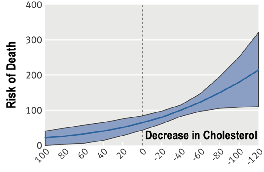
3/7) Cholesterol decreased substantially in the intervention group by 13.8% (−31.2 mg/dL); however, the survival curve suggested a trend toward higher all-cause mortality in the intervention (high–linoleic acid) group, as well as a dose-response relationship between greater cholesterol lowering and higher probability of death. Recall, this was a randomized controlled trial.
Additionally, focusing only on cardiovascular health, the intervention group also fared worse. Interestingly, they had an autopsy cohort. In this autopsy cohort, 41% (31/76) of participants in the intervention group had at least one myocardial infarct, whereas only 22% (16/73) of participants in the higher saturated fat, lower linoleic acid control group did.
They also conducted a meta-analysis of randomized trials as part of this publication that similarly found little support for the traditional diet–heart hypothesis.
The Hard Truth: Viagra Could Prevent Alzheimer’s Disease (🔗 in 5/5)
1/5) The talk of the town this last week has been the Nature paper showing that Lithium may help prevent Alzheimer’s disease. But what if another common (bedroom) compound could reduce Alzheimer’s risk by 69%? …
Viagra might prevent Alzheimer’s disease. And yes, I am serious. The paper I want to talk about was published in @NatureAging. The researchers began with an exploratory analysis, looking for predicted molecular interactions between existing drugs and pathways involved in Alzheimer’s disease.
Viagra (Sildenafil) stood up — darn it! — I meant, stood out.
2/5) Now, if Viagra truly reduces the risk of Alzheimer’s, we should expect to see that effect in large population datasets. And we do. The researchers performed multiple analyses on over 7 million individuals enrolled in Medicare Advantage insurance plans and found that Viagra use was associated with a 69% reduced risk of Alzheimer’s disease compared to non-users.
3/5) To reduce the chances of confounding variables, they also compared Viagra against other commonly prescribed medications — including diltiazem, glimepiride, losartan, and metformin. Every time, Viagra use was associated with a reduced Alzheimer’s risk compared to the comparison drug group.
2/8) Lithium: The Simplest Metal with Mysterious Power
Common and trace metals play critical roles in biology — think about zinc or iron. They’re essential for cell signaling, enzyme activation, and altering biochemical pathways. But the simplest metal of all, and one of the smallest elements on the periodic table, is lithium.
Despite its simplicity, lithium is biologically potent and mysterious.
At very high doses, lithium is a mood stabilizer commonly prescribed for bipolar disorder.
Fascinating, isn’t it? One of the simplest elements in the universe is a frontline treatment for severe mental illness.
Over the years, signals have emerged in the literature suggesting that lithium may protect against Alzheimer’s disease. For example, population studies have found that regions with higher levels of trace lithium in drinking water tend to have lower rates of Alzheimer’s.
Quoting from JAMA Psychiatry: “Exposure to higher long-term lithium levels in drinking water may be associated with a lower incidence of dementia.”
Coincidence? I doubt it.
3/8) We also know that lithium inhibits a key protein involved in Alzheimer’s pathology — Glycogen Synthase Kinase 3β (GSK3β).
The name doesn’t exactly reveal its mischief: GSK3β (also called tau kinase 1) adds phosphate groups to a protein called tau in the brain, which drives the formation of one of the core hallmarks of Alzheimer’s disease— phospho-tau neurofibrillary tangles.
As a matter of fact GSK3β promotes the formation of both neuropathological hallmarks of Alzheimer’s disease: (1) the infamous amyloid plaques and (2) phospho-tau neurofibrillary tangles.
Obesity Rewires Your Brain, Your Kidneys, and Your Blood Pressure (Full content🔗in 5/5; And 3/5 will freak you out 🫣)
1/5) For decades, we’ve been told to cut back on salt to control blood pressure.
The U.S. Dietary Guidelines still recommend limiting sodium intake to 2.3 grams per day.
But the science behind this advice is far from settled.
In fact, some long-term studies suggest the opposite: that lower sodium intake associates with higher blood pressure. (reference in letter, linked at the end).
And—curiously—people with obesity tend to be more salt-sensitive than lean individuals.
Why?
I promise, we’re going to unpack these questions today in the letter, and give you enough knowledge to terrify your cardiologist and woo your nephrologist.
2/5) Body Fat and Blood Pressure: A Complex Connection
Emerging research points to obesity as one culprit causing high blood pressure. Not just because of the extra weight, but because of what fat tissue does behind the scenes.
Let’s look at one such study—a remarkable tour de force published in @Cell_Metabolism ... In this study, researchers fed mice a high-sugar, high-fat, obesogenic diet. Blood pressure didn’t rise right away; it only increased after the mice had become obese. But something else happened first…
3/5) The high-sugar, high-fat diet caused obesity, which contributed to a thickening of vessels around the hypothalamus—something clearly visible below.
🧠The microscope images on the left are more zoomed-out and show how the high-sugar, high-fat diet increases the overall density of vessels around the hypothalamus at the base of the brain.
🧠The images on the right are more zoomed-in, showing blood vessels under a powerful electron microscope. What’s shown is a significant thickening—highlighted in the blue insets—of the basal membranes of the vessels.
If that didn’t totally make sense, the takeaway is simple: the high-sugar, high-fat diet caused obesity, an increase in leptin, and a significant remodeling of vessels around the brain’s hypothalamus. And—importantly—this vascular remodeling coincided with rising body fat and leptin levels and preceded the rise in blood pressure.