
The JVP exam has been with us since 1930 when Sir Thomas Lewis first proposed this exceptionally clever technique. A #pocus thread. 
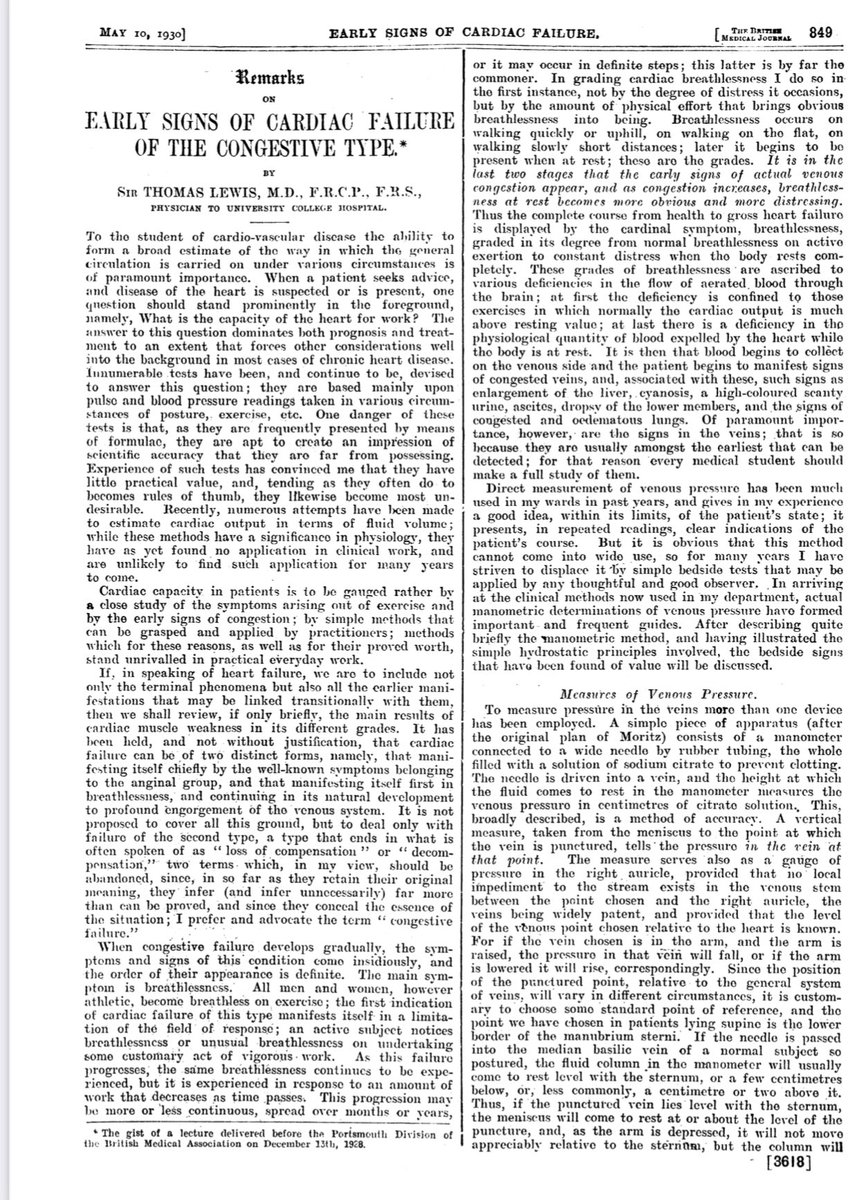
He knew that in heart failure there in “congestion of the veins” and when they become engorged can be detected on the neck.
Here is a phenomenal example in a very thin patient with prominent veins.
However most patients do not have such a visible IJ. Whether from a thick neck, obesity, large beard or otherwise, the JVP can be invisible in many patients. In these patients it cannot be determined if the JVP is low and below the clavicle, so high that it resides above the jaw
or present in the neck and cannot be seen. This is where #pocus can be very useful
In that same thin patient this is their IJ at the top and his carotid in the bottom right. Note the distinction of the IJ with minimal pulsations.
In the longitudinal view you can see the carotid below the IJ. The meniscus of the IJ takes a triangular shape
Note the pulsations returning. And when he breathes in the IJ expands. This is where the pulsations are most prominent, the sonographic version of the top of the blood column. It is from this point when you would measure the height of the blood column.
And with #pocus “JVP is ALWAYS appreciated.” This and much more in our @The_POCUS_Book coming out very VERY soon. #cardiotwitter #foamed #meded #cardiology #echofirst
• • •
Missing some Tweet in this thread? You can try to
force a refresh


