This study from #Singapore compares the immune characteristics of 55 patients with vaccine breakthrough #SARSCoV2 infection and 86 uninfected vaccinated close contacts. 1/
Antibody levels, including neutralizing antibodies, were similar in vaccine breakthrough patients and close contacts. 2/
Memory B cell levels, as assessed by B cell ELISpot, were lower in vaccine breakthrough patients than close contacts. 3/
T cell profiles were broadly similar across vaccine breakthrough patients and close contacts. 4/
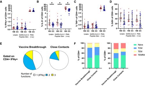
The cytokine profile of vaccine breakthrough patients was similar to uninfected vaccinated individuals, with lower inflammatory profile compared to unvaccinated individuals with primary infection. 5/
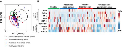
Conclusions:
1-These results highlight the potential role of memory B cells in protection from Delta vaccine breakthrough infection.
2-The results suggest that memory B cell levels may be a correlate of protection against Delta variant infection in vaccinated populations 6/
3-If so, this will be useful for determining the level of susceptibility in a population. It will also be useful in the design of future vaccines or vaccine boosters. 7/
Could even mild COVID-19 leave a lasting imprint on the brain?
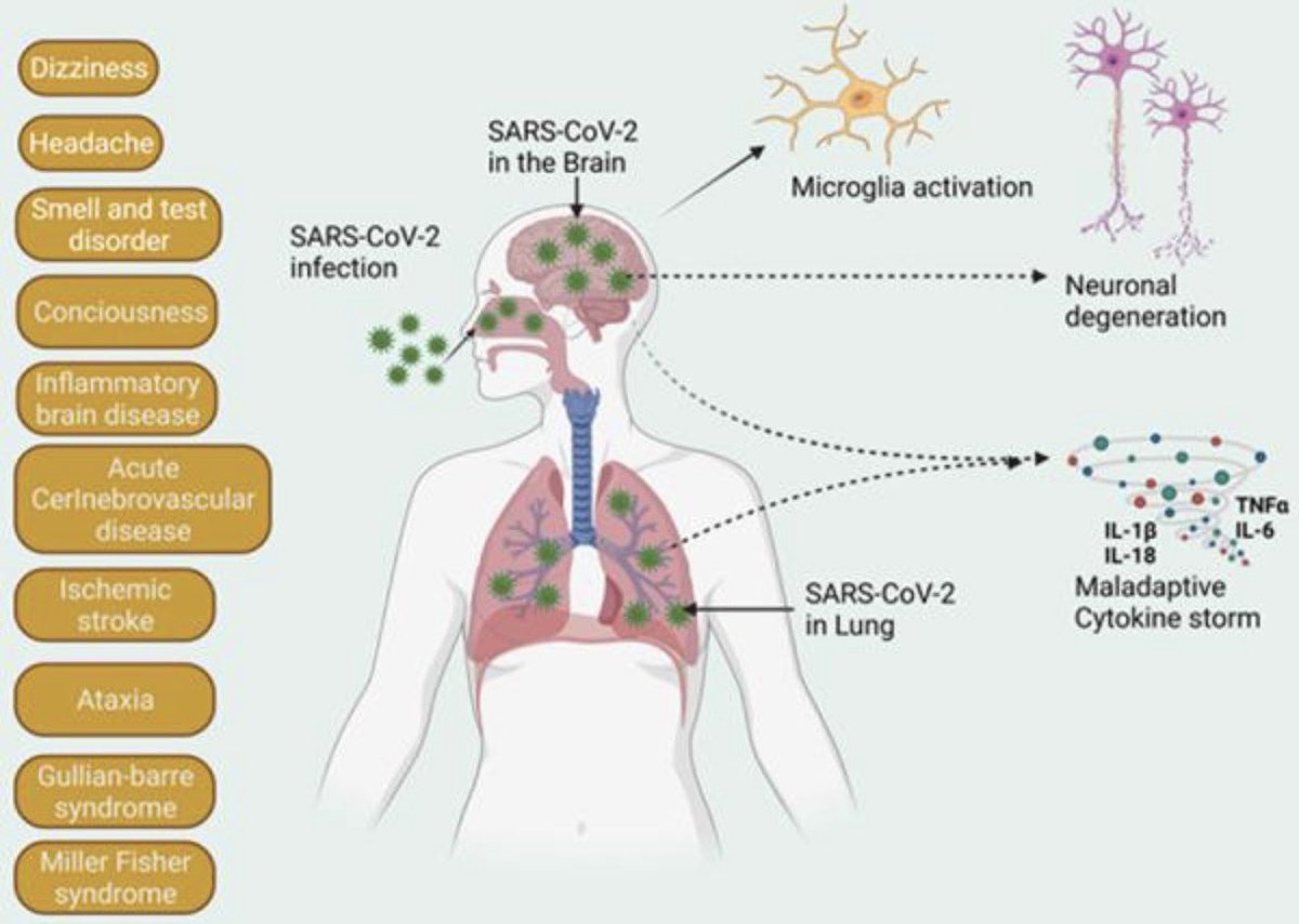
➡️ Using a non-human primate model, researchers found that mild SARS-CoV-2 infection triggered weeks of persistent neuroinflammation, with dopaminergic neuron loss in the substantia nigra—the brain region most vulnerable in Parkinson’s disease.
Markers of neuronal injury remained elevated in both blood and cerebrospinal fluid. 1/
Even more striking, the CSF proteome shifted toward patterns seen in neurodegenerative diseases, including alterations in proteins linked to Alzheimer’s disease, suggesting that SARS-CoV-2 may transiently activate biological pathways shared with neurodegeneration. 2/
The work reports that in a non-human primate model, even mild Delta variant infection resulted in:
➡️ Persistent neuroinflammation in the hippocampus and pons.
➡️ Dopaminergic neuron loss in the substantia nigra.
➡️ Elevated neurofilament light chain (NfL) in blood and CSF. 3/
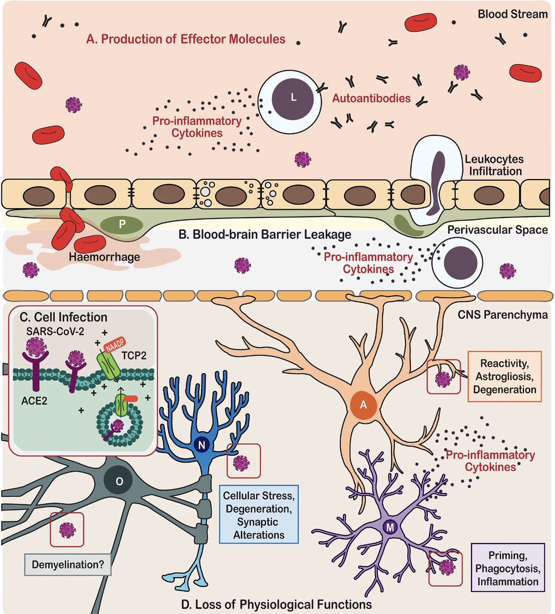
What if #LongCOVID brain fog doesn’t start in the brain—but in its protective coverings?
➡️ A new review proposes that the meninges, the membranes surrounding the brain, may serve as a reservoir for persistent SARS-CoV-2 Spike protein.
➡️ These membranes are not passive coverings—they are highly active immune organs packed with mast cells, blood vessels, and lymphatic channels. 1/
The implication is profound: LongCOVID may originate from inflammation around the brain rather than within neurons themselves.
➡️The paradigm may be shifting—from viewing LongCOVID as a purely neuronal disorder to recognizing it as a disease of neuroimmune interactions at the brain’s borders. 2/
Meet the brain’s overlooked troublemakers: mast cells.
➡️ The review suggests that persistent Spike protein may chronically activate meningeal mast cells, which release a barrage of histamine, cytokines, proteases, and other vasoactive mediators. 3/
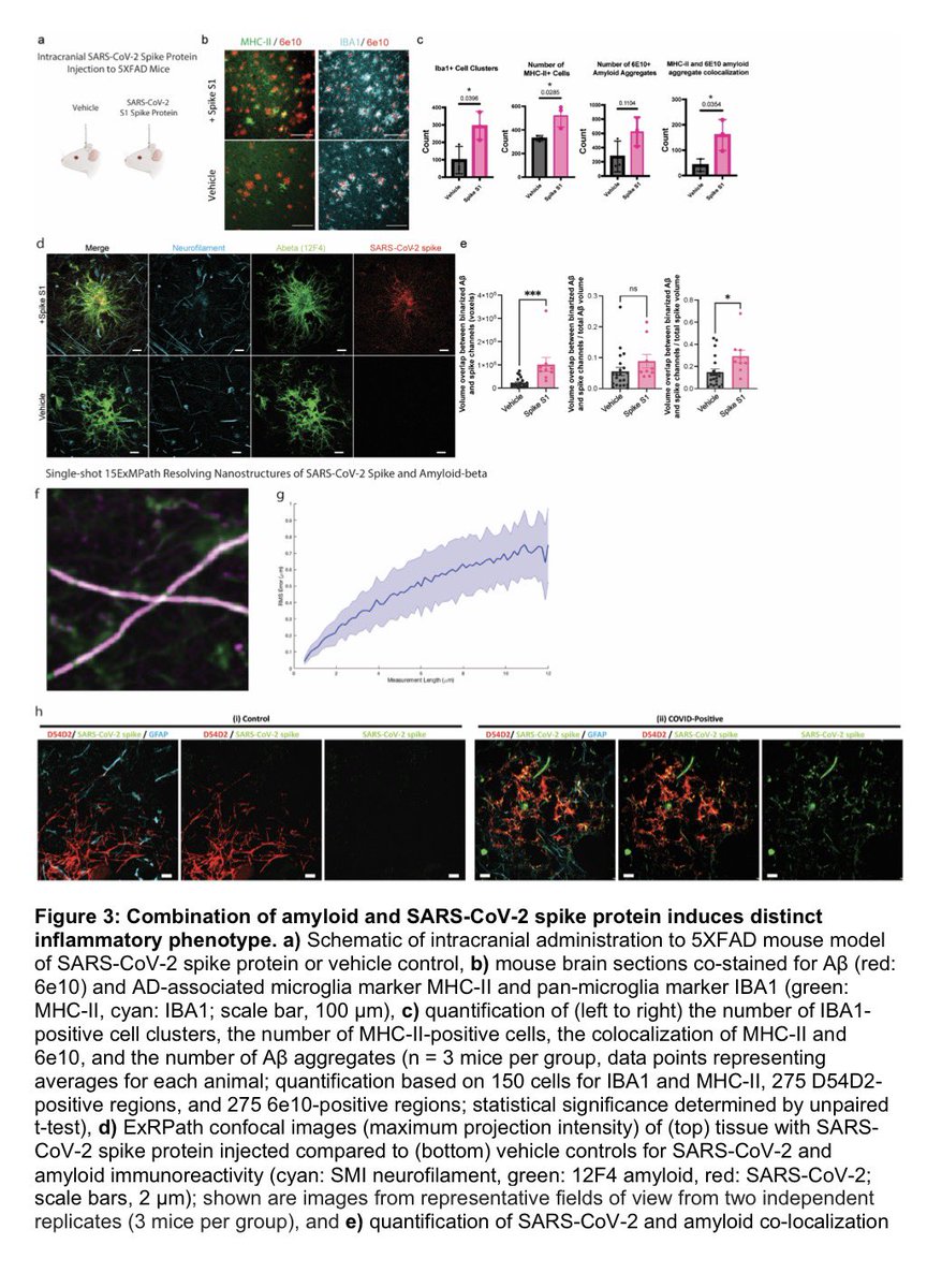
SARS-CoV-2 spike protein may directly amplify brain inflammation.
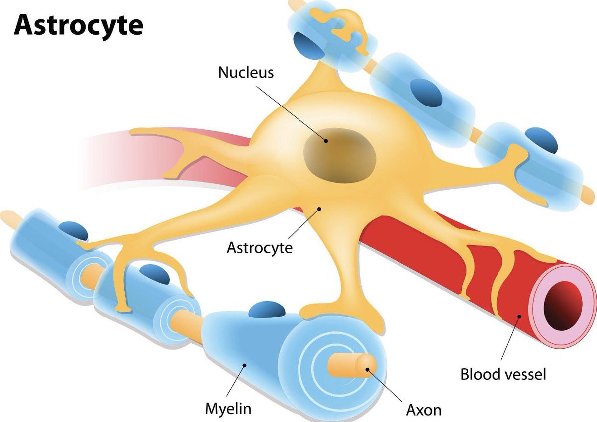
➡️ Researchers found that spike proteins can colocalize with amyloid-β (Aβ) and trigger distinct inflammatory responses in microglia — the brain’s immune cells.
➡️ This raises important questions about potential long-term neurodegenerative consequences of COVID-19. 1/
Researchers developed advanced “expansion microscopy” techniques that physically enlarge human brain tissue, allowing scientists to see disease-related structures at near-nanoscale resolution using ordinary microscopes. 2/
Applying this method to brains from some COVID-19 patients revealed tiny amyloid-like protein clusters closely associated with SARS-CoV-2 particles in a small subset of cases, suggesting a possible link between COVID-19, neuroinflammation, and abnormal protein aggregation in the brain.
The study highlights how ultra-high-resolution imaging could uncover previously hidden mechanisms of neurological disease. 3/
👉 The lungs may remain biologically altered long after acute infection resolves. 1/
A new review highlights how persistent immune activation in LongCOVID may lead to:
• Fibrosis-like lung changes
• Endothelial dysfunction
• Microvascular injury
• Ongoing respiratory symptoms
COVID may end clinically—but not biologically.
#LongCOVID #Pulmonology 2/
LongCOVID respiratory sequelae may result from a “perfect storm” of:
COVID-19 may be, in part, a mitochondrial disease.
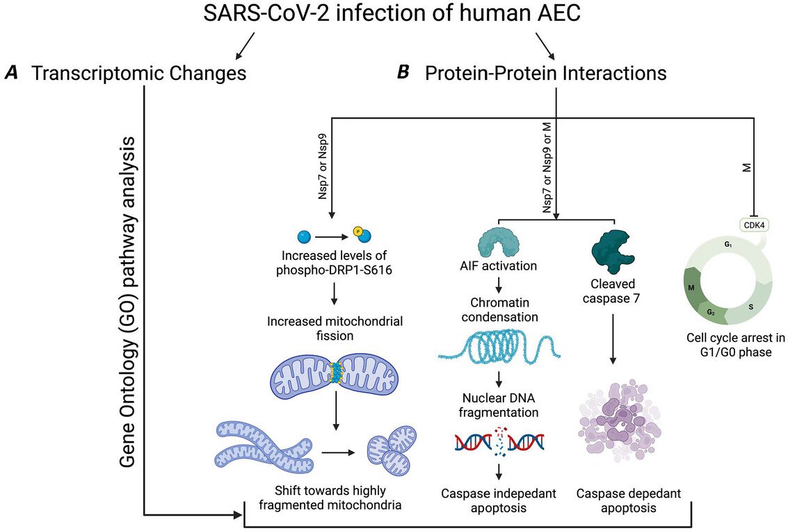
➡️ A Cambridge review shows SARS-CoV-2 disrupts mitochondrial function in lung cells—driving inflammation and worsening pneumonia.
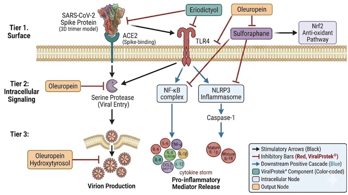
➡️ Emerging studies suggest even after the active infection is resolved, residual viral proteins, particularly SARS-CoV-2 spike protein, may linger and continue to cause damage to the mitochondria by increasing oxidative stress and disrupting energy metabolism, offering a plausible mechanism for #LongCOVID. 1/
H/T: @CatchTheBaby
COVID-19 is not just viral—it’s metabolic.
SARS-CoV-2 hijacks mitochondria →
↓ Energy production
↑ Inflammatory signaling
A key pathway worsening lung injury. 2/
Mitochondria may link acute COVID → #LongCOVID.
Viral disruption of mitochondrial function can persist, sustaining oxidative stress and immune dysregulation even after infection. 3/