Aaron Rutman, MD Profile picture
May 24 22 tweets 12 min read
Pterygopalatine fossa🧵- inspired by ?s from med students in neuroanatomy lab & a resident w/ case of perineural tumor spread on same day! #meded #FOAMrad #medtwitter #medstudents #radiology #neurorad #HNrad #radres #neurology #ENT #temporalbone #neurosurgery #neuroanatomy
1/22
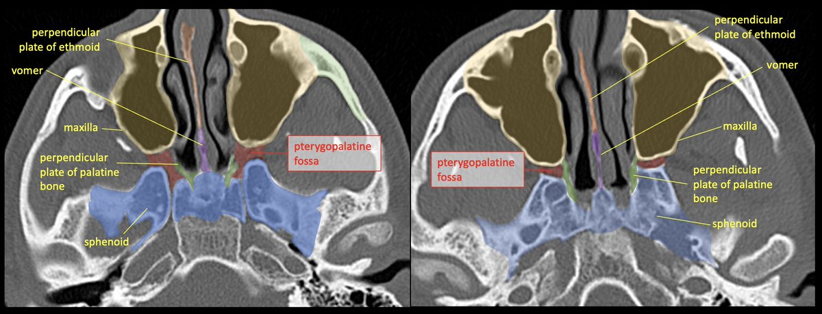
The PTERYGOPALATINE FOSSA (PPF) is a space deep in the face/skull base, bordered anteriorly by the maxilla (max sinus), posteriorly by the pterygoid base of the sphenoid, and medially by the perpendicular plate of the palatine bone. 2/22
It’s best to think of the PPF as a crossroads/intersection. Think about the roads that lead to and from it, and the cast of characters that pass through.

Some like to simplify/visualize the PPF as a cube or inverted pyramid.

3/22
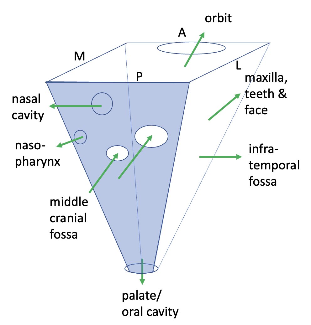
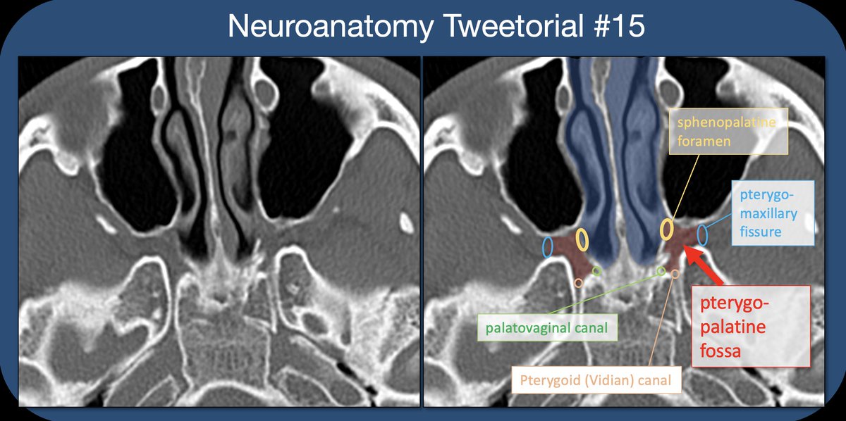
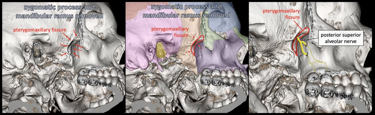
The PPF is accessed from laterally via the PTERYGOMAXILLARY FISSURE (PMF), an open crevasse between the pterygoid base of the sphenoid and the post maxilla. On 3D images, It can be seen deep to the zygomatic process.
4/22
The PMF is the lateral exit route from PPF; the POST-SUP ALVEOLAR NERVE branches from the INFRAORBITAL nerve & travels along the lateral margin of maxilla before diving into the wall to innervate the maxillary molars (also innervates max sinus and post oral mucosa).
5/22
Within the PPF lives the PTERYGOPALATINE GANGLION (PPG) which contains post-synaptic parasympathetic cell bodies-->lacrimal gland and sinus/nasal/oral mucosa. Many other sympathetic and somatic nerves travel through the ganglion without synapsing.
6/22
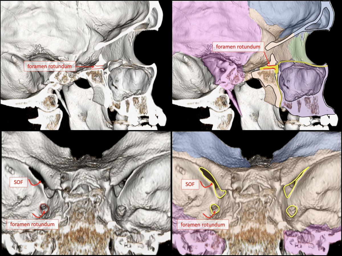
There are two canals which travel in the AP plane through the sphenoid bone, and transmit nerves/vessels from the middle cranial fossa to the PPF: FORAMEN ROTUNDUM and the PTERYGOID (VIDIAN) CANAL. The pterygoid canal is more inf and medial.
7/22
The FORAMEN ROTUNDUM transmits V2, the MAXILLARY NERVE, from the middle cranial fossa to the PPF in the AP direction. The Maxillary nerve (V2) contains mainly sensory information from the mid-face.
8/22
The PTERYGOID CANAL transmits the NERVE OF THE PTERYGOID CANAL through the sphenoid from mid cranial fossa (ant to foramen lacerum) to the PPF. Contains preganglionic parasympathetic (from gr sup petrosal n) and post-ganglionic sympathetic (deep petrosal n) from ICA plexus.
9/22
Anterior/superior exit route from the PPF is the INFERIOR ORBITAL FISSURE (IOF). The IOF is formed by the sphenoid and maxilla, connecting the PPF to the floor of the orbital apex.
10/22
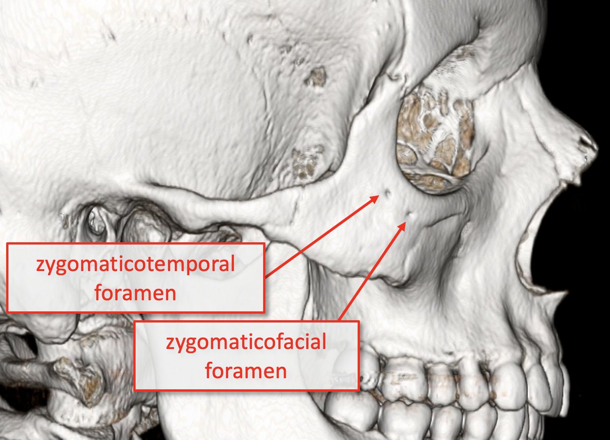
The IOF transmits the ZYGOMATIC and INFRAORBITAL NERVES (V2 branches); ZYGOMATIC n. supplies sensory info from skin over zygomatic/temporal bones (zygomaticotemporal and zygomaticofacial branches) & transmits postganglionic parasympathetic fibers to lacrimal nerve/gland.
11/22
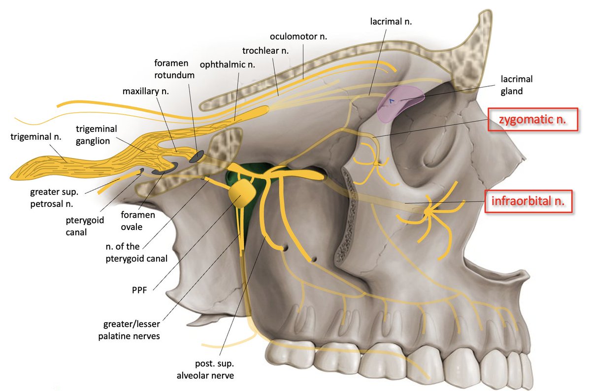
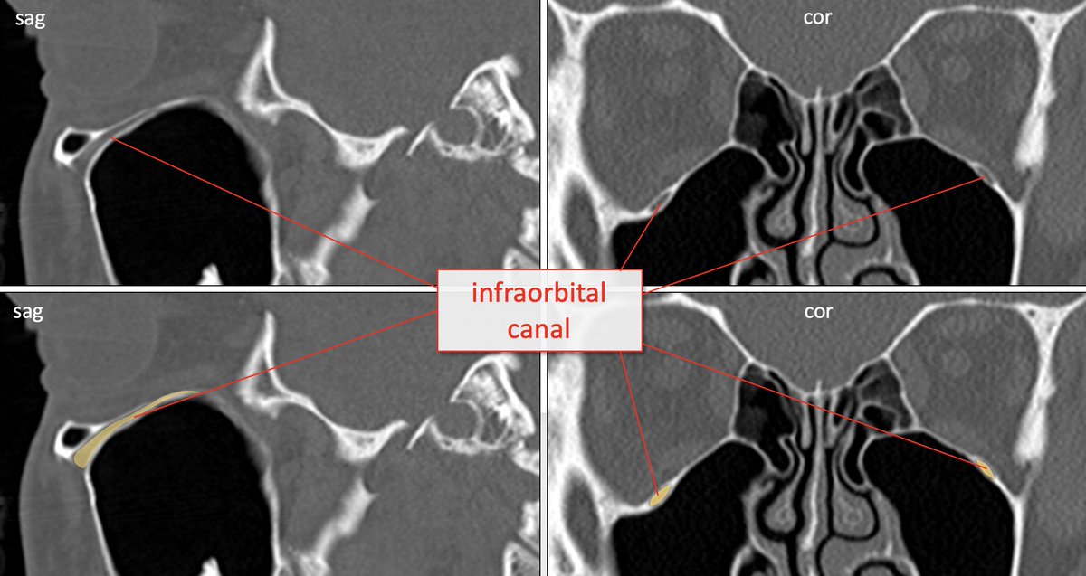
The INFRAORBITAL NERVE branches from V2 in the PPF➡️goes through IOF➡️extends anteriorly➡️ infraorbital groove/canal at orbital floor➡️exits INFRAORBITAL FORAMEN of the maxilla; supplies sensory to skin of cheek/lower eyelid.
12/22
The middle and ant sup alveolar nerves variably branch from the infraorbital nerve to supply sensory information from the maxillary teeth.
13/22
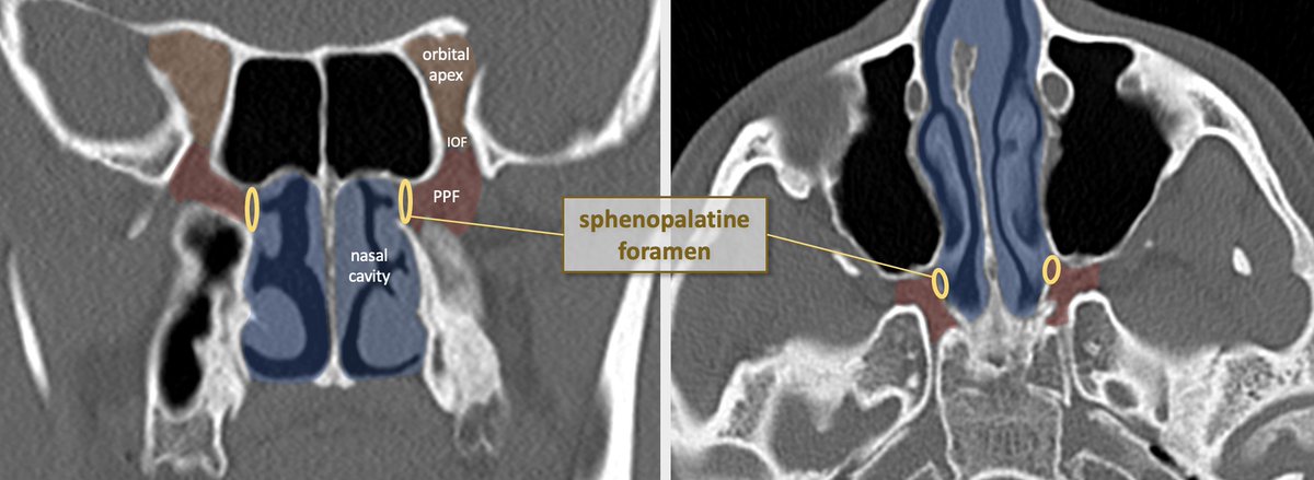
The medial exit route of the PPF leads to the nasal cavity via the SPHENOPALATINE FORAMEN which is formed by the palatine and sphenoid bones. It transmits the NASOPALATINE NERVE, posterior superior nasal nerves, and sphenopalatine artery/vein.
14/22
The NASOPALTINE NERVE branches from V2 in PPF (just after from f. rotundum)➡️traverses sphenopalatine foramen➡️courses within nasal septum anteroinferiorly➡️goes through INCISIVE FORAMEN of the maxillary palate (somatic sensory & autonomic supply to nasal mucosa).
15/22
The POST SUP NASAL NERVES also branch from V2 in PPF, traverse sphenopalatine foramen, and supply posterior nasal septum and lateral nasal wall.
16/22
Posteriorly and medially, the PHARYNGEAL NERVE (branch of V2) exits the PPF to the nasopharynx via the PALATOVAGINAL CANAL. It contains somatic sensory info and autonomic motor supplying the sup nasopharyngeal mucosa.
17/22
Inferiorly, the GREATER and LESSER PALATINE NERVES exit the PPF via the GREATER and LESSER PALATINE FORAMINA to the greater/lesser palatine canals:
18/22
The greater palatine foramen is located at the lateral suture between the maxillary palate and palatine bone; lesser canals are within the horizontal plate of the palatine bone. Nerves contain somatic sensory and autonomic innervation to the palate and oral cavity.
19/22
Thats the PPF! Important because so many functions, (including including midface sensation, sinonasal and pharyngeal sensation, pharyngeal/sinonasal mucus and saliva production, and lacrimation) all pass through this small anatomic space.
20/22
Importantly, mucosal tumors such as squamous cell CA and adenoid cystic CA love to spread along the nerves (perineural spread). Staging of pharynx, palate, sinus, salivary gland tumors requires knowledge of nerve pathways; knowing the PPF is imperative!
21/22
Clinical Case: adenoid cystic CA of the left palate, with associated perineural spread involving the palatine nerves, PPF, post/sup alveolar n, infraorbital n, maxillary n, mandibular n, and Meckel's cave.
22/22

• • •

Missing some Tweet in this thread? You can try to force a refresh
 

Keep Current with Aaron Rutman, MD

Aaron Rutman, MD Profile picture

Stay in touch and get notified when new unrolls are available from this author!

Read all threads

This Thread may be Removed Anytime!

PDF

Twitter may remove this content at anytime! Save it as PDF for later use!

Try unrolling a thread yourself!

how to unroll video
  1. Follow @ThreadReaderApp to mention us!

  2. From a Twitter thread mention us with a keyword "unroll"
@threadreaderapp unroll

Practice here first or read more on our help page!

More from @aaronrutman

Mar 6, 2021
Neuroanatomy TOTD #15🧵
The inner ear #tweetorial--it packs a large functional punch for its small size-strap in!
#meded #FOAMed #FOAMrad #medtwitter #medstudents #radiology #neurorad #HNrad #radres #neurology #ENT #temporalbone #neurosurgery #neuroanatomy #neuroanatomyTOTD
1/24
To evaluate the t-bone, best to compartmentalize--external/middle/inner ear (IE). See previous #tweetorial of the ME. The IE is difficult as most structures are obliquely oriented (at different obliquities!)-and can be hard to see on standard views. 2/24
IE communicates with ME via oval&round windows (which allow for transmission&dissipation of sonic vibrations). IE houses sensory organs for hearing/balance/sensing motion. The cochlear&vestibular nerves (CNVIII) transmit signals to the brain via the int auditory canal (IAC). 3/24
Read 25 tweets
Jan 11, 2021
Neuroanatomy TOTD #14🧵
Got some requests to do one of the trickiest areas of human anatomy, the #temporalbone. So many named structures! #meded #FOAMed #FOAMrad #medtwitter #medstudents #radiology #neurorad #radres #neurosurgery #neuroanatomy #ENT #otolaryngology

1/21 Image
Whether learning t-bone anatomy as a medical student or evaluating a CT of the t-bone as a radres, it’s best to compartmentalize into external, middle, and inner ear. This thread🧵is on the middle ear: Inner ear to follow, some day:)

2/21 Image
The external ear extends from the external meatus to the TM. The TM should be thin and *almost* imperceptible on CT. Thickened and retracted TM suggests prior pathology (usually otitis media) and scarring.

3/21 Image
Read 22 tweets
Dec 2, 2020
Neuroanatomy TOTD #12

The green structure is the amygdala (amygdaloid body) and the yellow structure is the stria terminalis (ST).

1/18

#meded #FOAMed #FOAMrad #medtwitter #medstudents #radiology #neurorad #radres #neurology #neurosurgery #neuroanatomy #neuroanatomyTOTD
Time for a deep dive into limbic networks. Bear with me—this is a fun subject. I got carried away preparing slides—it’s hard to know when to stop!

2/18
The amygdala is an almond-shaped collection of gray nuclei/subnuclei deep to the uncus and ant to hippocampus in the med temporal lobe. Involved in multiple functions: memory modulation, emotional learning and responses, +important connections w/ the olfactory bulb/cortex.

3/18
Read 18 tweets
Nov 15, 2020
Neuroanatomy TOTD #11🧵
➡️intracanalicular facial nerve. IAC cross section➡️4 nerves; ant-sup➡️facial, ant-inf➡️cochlear (7-up/coke-down). Post nerves➡️sup&inf vestibular #medtwitter #meded #FOAMed #FOAMrad #neurorad #radiology #radres #neurology #neurosurgery #neuroanatomy
1/11
Facial motor nucleus is in the pontine tegmentum; axons loop dorsally around the abducens nucleus, then course anterolaterally, exiting the brainstem at the CPA.
2/11
The nervus intermedius (NI) is composed of preganglionic parasympathetic fibers w/cell bodies in the sup salivatory nucleus, taste fibers w/cell bodies in the nucleus solitarius, and somatic sensory input from EAC/external ear, w/cell bodies in the spinal nucleus of CNV.

3/11
Read 11 tweets
Oct 30, 2020
Neuroanatomy TOTD #10🧵 
1/5 Small gray matter structure at the junction of the thalamus and midbrain is the subthalamic nucleus (STN).

#meded #FOAMed #FOAMrad #medtwitter #medstudents #radiology #neurorad #radres #neurology #neurosurgery #neuroanatomy #neuroanatomyTOTD
2/5...The STN is functionally a node within the basal ganglia (BG) INDIRECT LOOP. STN contains excitatory glutaminergic neurons➞output to the GABA neurons of GPi, which in turn have inhibitory effect on thalamic outputs to the motor cortex.
3/5...Loss of nigrostriatal input in Parkinsons Dz➞increased inhibitory output from GPi➞decreased thalamic stimulation of the motor cortex (through both direct and indirect loop circuits). Makes sense that DBS treatments were initially directed at disrupting activity in GPi.
Read 5 tweets
Oct 23, 2020
Neuroanatomy TOTD #9🧵
1/6 The trigeminal n. courses anteriorly➡️prepontine cistern➡️into Meckel’s cave (green), which lies at the medial floor of middle cranial fossa at the petrous apex.
#meded #FOAMed #FOAMrad #neurorad #neurology #neurosurgery #neuroanatomy #neuroanatomyTOTD Image
2/6...Meckel’s cave (MC) is open to the subarachnoid space at its posterior margin (and is therefore filled with CSF). The trigeminal (Gasserion) ganglion lives in MC. Superiomedial to MC (and sharing a dural border), is the cavernous sinus. #radres #neurorad Image
3/6...Coursing anteriorly, the V1 and V2 branches of CNV exit MC to travel within the lateral wall of the cavernous sinus. V3 courses inferiorly to exit the middle cranial fossa through foramen ovale, without involving the cavernous sinus. Image
Read 6 tweets

Did Thread Reader help you today?

Support us! We are indie developers!


This site is made by just two indie developers on a laptop doing marketing, support and development! Read more about the story.

Become a Premium Member ($3/month or $30/year) and get exclusive features!

Become Premium

Don't want to be a Premium member but still want to support us?

Make a small donation by buying us coffee ($5) or help with server cost ($10)

Donate via Paypal

Or Donate anonymously using crypto!

Ethereum

0xfe58350B80634f60Fa6Dc149a72b4DFbc17D341E copy

Bitcoin

3ATGMxNzCUFzxpMCHL5sWSt4DVtS8UqXpi copy

Thank you for your support!

Follow Us on Twitter!

:(