Lea Alhilali, MD Profile picture
Jun 3, 2022 7 tweets 5 min read Read on X
1/”Tell me where it hurts.” How back pain radiates can tell you where the lesion is—if you know where to look!
A 🧵about how to remember lumbar radicular pain distributions. #medstudenttwitter #medtwitter #radres #FOAMed #FOAMrad #neurorad #Meded #backpain #spine #Neurosurgery Image
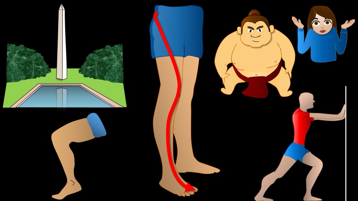
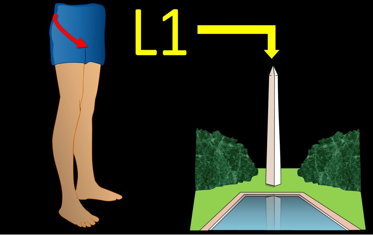
2/Let’s start with L1. L1 radiates to the groin. I remember that b/c the number 1 is, well, um…phallic. So the phallic number 1 radiates to the groin. Image
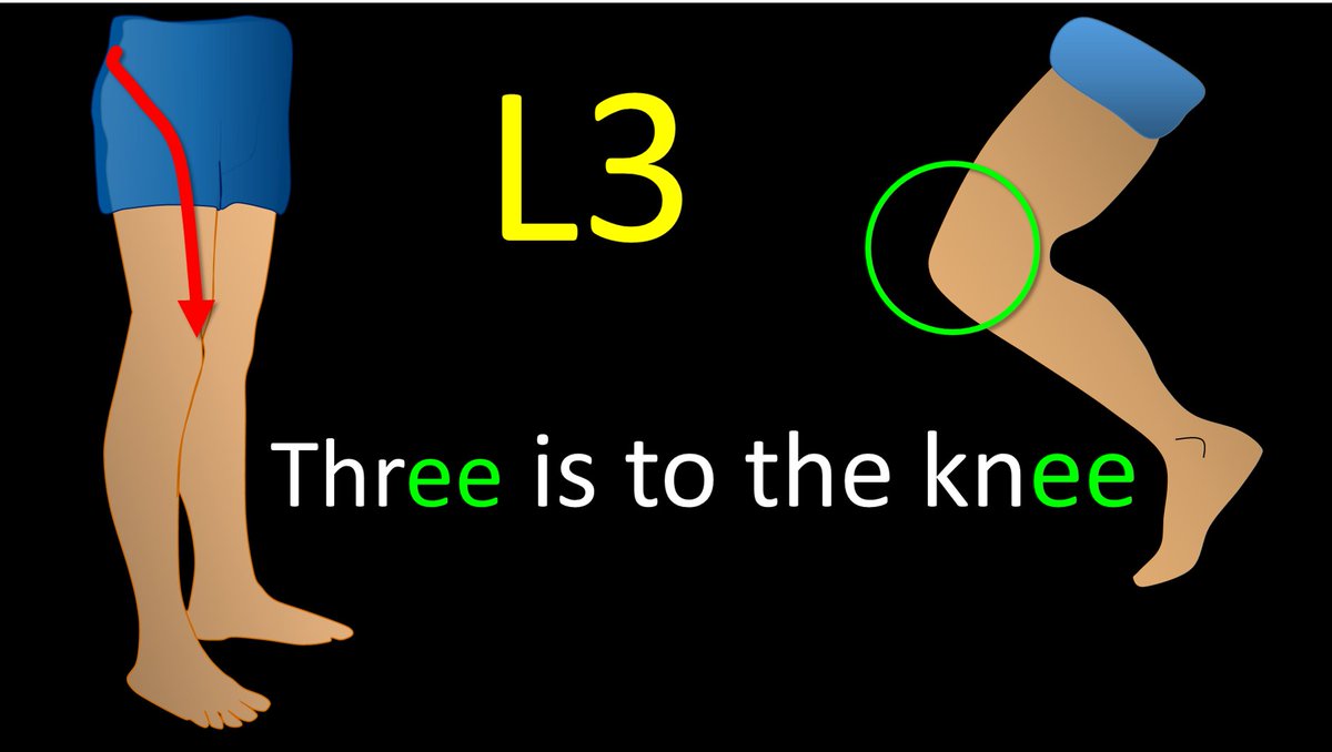
3/Let’s skip to L3 for a second. I remember L3 is to the knee—easy, it rhymes! Image
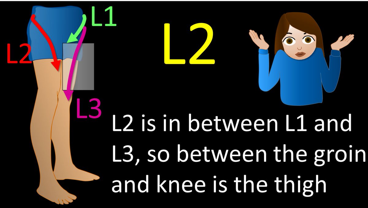
4/Ok, back to L2. Two is the number between 1 and 3, so the distribution of L2 is between the distributions of L1 and L3—and between the groin and knee is the thigh. L2 radiates to the thigh. It’s not the catchiest way to remember it, but it works. Image
5/L4 radiates to the calf. I remember this bc the number 4 looks like the calf, with the top part of the 4 looking like a bulging gastroc & the bottom part of the four is the rest of the calf connecting to the ankle. Don’t we all wish we had bulging gastrocs like the number 4! Image
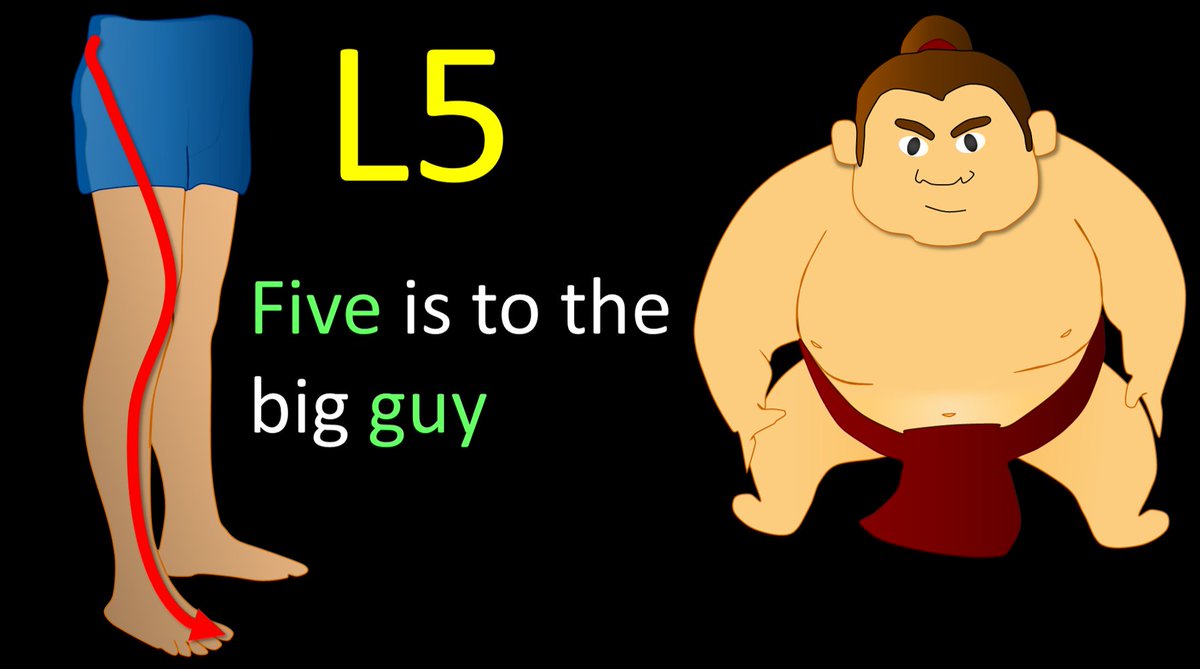
6/L5 radiates to the big toe. So I have the little rhyme “Five is to the big guy!” L5 is also foot drop. So I remember big guys are heavy, and heavy gravity = drop. If I hear the history “foot drop,” I never stop looking until I have traced out the entire L5 nerve root. Image
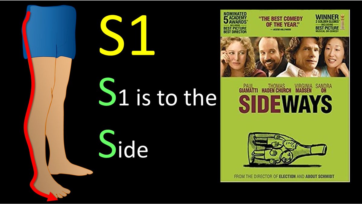
7/Finally, S1 radiates to the side of the foot. I remember this bc both S1 & Side start w/S.
So now you know where in the lumbar spine to look when a patient says the pain radiates down the leg & hopefully remembering the lumbar radicular distributions won’t cause you any pain! Image

• • •

Missing some Tweet in this thread? You can try to force a refresh
 

Keep Current with Lea Alhilali, MD

Lea Alhilali, MD Profile picture

Stay in touch and get notified when new unrolls are available from this author!

Read all threads

This Thread may be Removed Anytime!

PDF

Twitter may remove this content at anytime! Save it as PDF for later use!

Try unrolling a thread yourself!

how to unroll video
  1. Follow @ThreadReaderApp to mention us!

  2. From a Twitter thread mention us with a keyword "unroll"
@threadreaderapp unroll

Practice here first or read more on our help page!

More from @teachplaygrub

Jul 25
1/Time to go with the flow!

Hoping no one notices you don’t know the anatomy of internal carotid (ICA)?

Do you say “carotid siphon” & hope no one asks for more detail?

Here’s a thread to help you siphon off some information about ICA anatomy! Image
2/ICA is like a staircase—winding up through important anatomic regions like a staircase winding up to each floor Lobby is the neck.

First floor is skullbase/carotid canal. Next it stops at the cavernous sinus, before finally reaching the rooftop balcony of the intradural space.Image
3/ICA is divided into numbered segments based on landmarks that denote transitions on its way up the floors.

C1 is in the lobby or neck.

You can remember this b/c the number 1 looks elongated & straight like a neck. Image
Read 10 tweets
Jul 23
1/My hardest thread yet! Are you up for the challenge?

How stroke perfusion imaging works!

Ever wonder why it’s Tmax & not Tmin?

Do you not question & let RAPID read the perfusion for you? Not anymore! Image
2/Perfusion imaging is based on one principle: When you inject CT or MR intravenous contrast, the contrast flows w/blood & so contrast can be a surrogate marker for blood.

This is key, b/c we can track contrast—it changes CT density or MR signal so we can see where it goes. Image
3/So if we can track how contrast gets to the tissue (by changes in CT density or MR signal), then we can approximate how BLOOD is getting to the tissue.

And how much blood is getting to the tissue is what perfusion imaging is all about. Image
Read 18 tweets
Jul 21
1/Do you know all the aspects of, well, ASPECTS?

Many know the anterior circulation stroke scoring system—but posterior circulation (pc) ASPECTS is often left behind

25% of infarcts are posterior circulation

Do you know pc-ASPECTS?!

Here’s how to remember pc-ASPECTS! Image
2/Many know anterior circulation ASPECTS.

It uses a 10-point scoring system to semi-quantitation the amount of the MCA territory infarcted on non-contrast head CT

If you need a review: here’s my thread on ASPECTS: Image
3/But it’s only useful for the anterior circulation.

Posterior circulation accounts for ~25% of infarcts.

Even w/recanalization, many of these pts do poorly bc of the extent of already infarcted tissue.

So there’s a need to quantitate the amount of infarcted tissue in these ptsImage
Read 12 tweets
Jul 2
1/The medulla is anything but DULL!

Does seeing an infarct in the medulla cause your heart to skip a beat?

Does medullary anatomy send you into respiratory arrest?

Never fear, here is a thread on the major medullary syndromes! Image
2/The medulla is like a toll road.

Everything going down into the cord must pass through the medulla & everything from the cord going back up to the brain must too.

That’s a lot of tracts for a very small territory. Luckily you don’t need to know every tract Image
3/Medulla has 4 main vascular territories, spread out like a fan: anteromedial, anterolateral, lateral, and posterior.

You don’t need to remember their names, just the territory they cover—and I’ll show you how Image
Read 18 tweets
Jun 30
1/Time is brain! But what time is it?

If you don’t know the time of stroke onset, are you able to deduce it from imaging?

Here’s a thread to help you date a stroke on MRI! Image
2/Strokes evolve, or grow old, the same way people evolve or grow old.

The appearance of stroke on imaging mirrors the life stages of a person—you just have to change days for a stroke into years for a person

So 15 day old stroke has features of a 15 year old person, etc. Image
3/Initially (less than 4-6 hrs), the only finding is restriction (brightness) on diffusion imaging (DWI).

You can remember this bc in the first few months, a baby does nothing but be swaddled or restricted. So early/newly born stroke is like a baby, only restricted Image
Read 10 tweets
Jun 27
1/”I LOVE spinal cord syndromes!” is a phrase that has NEVER, EVER been said by anyone.

Do you become paralyzed when you see cord signal abnormality?

Never fear—here is a thread on all the incomplete spinal cord syndromes to get you moving again! Image
2/Spinal cord anatomy can be complex. On imaging, we can see the ant & post nerve roots. We can also see the gray & white matter. Hidden w/in the white matter, however, are numerous efferent & afferent tracts—enough to make your head spin. Image
3/Lucky for you, for the incomplete cord syndromes, all you need to know is gray matter & 3 main tracts. Anterolaterally, spinothalamic tract (pain & temp). Posteriorly, dorsal columns (vibration, proprioception, & light touch), & next to it, corticospinal tracts—providing motor Image
Read 20 tweets

Did Thread Reader help you today?

Support us! We are indie developers!


This site is made by just two indie developers on a laptop doing marketing, support and development! Read more about the story.

Become a Premium Member ($3/month or $30/year) and get exclusive features!

Become Premium

Don't want to be a Premium member but still want to support us?

Make a small donation by buying us coffee ($5) or help with server cost ($10)

Donate via Paypal

Or Donate anonymously using crypto!

Ethereum

0xfe58350B80634f60Fa6Dc149a72b4DFbc17D341E copy

Bitcoin

3ATGMxNzCUFzxpMCHL5sWSt4DVtS8UqXpi copy

Thank you for your support!

Follow Us!

:(