Lea Alhilali, MD Profile picture
Jun 6, 2022 6 tweets 4 min read Read on X
1/Even attendings have to use google sometimes! Never be afraid to do whatever it takes to get the right diagnosis
A🧵about an interesting spine case that I gasped, “What the…” when I opened it #medtwitter #radres #FOAMed #FOAMrad #neurorad #Meded #backpain #spine #Neurosurgery Image
2/Older pt w/lower extremity numbness. MRI showed intradural, extra-axial rounded lesions in the lower thoracic region that avidly enhanced on post-contrast imaging—like gang busters enhancement Image
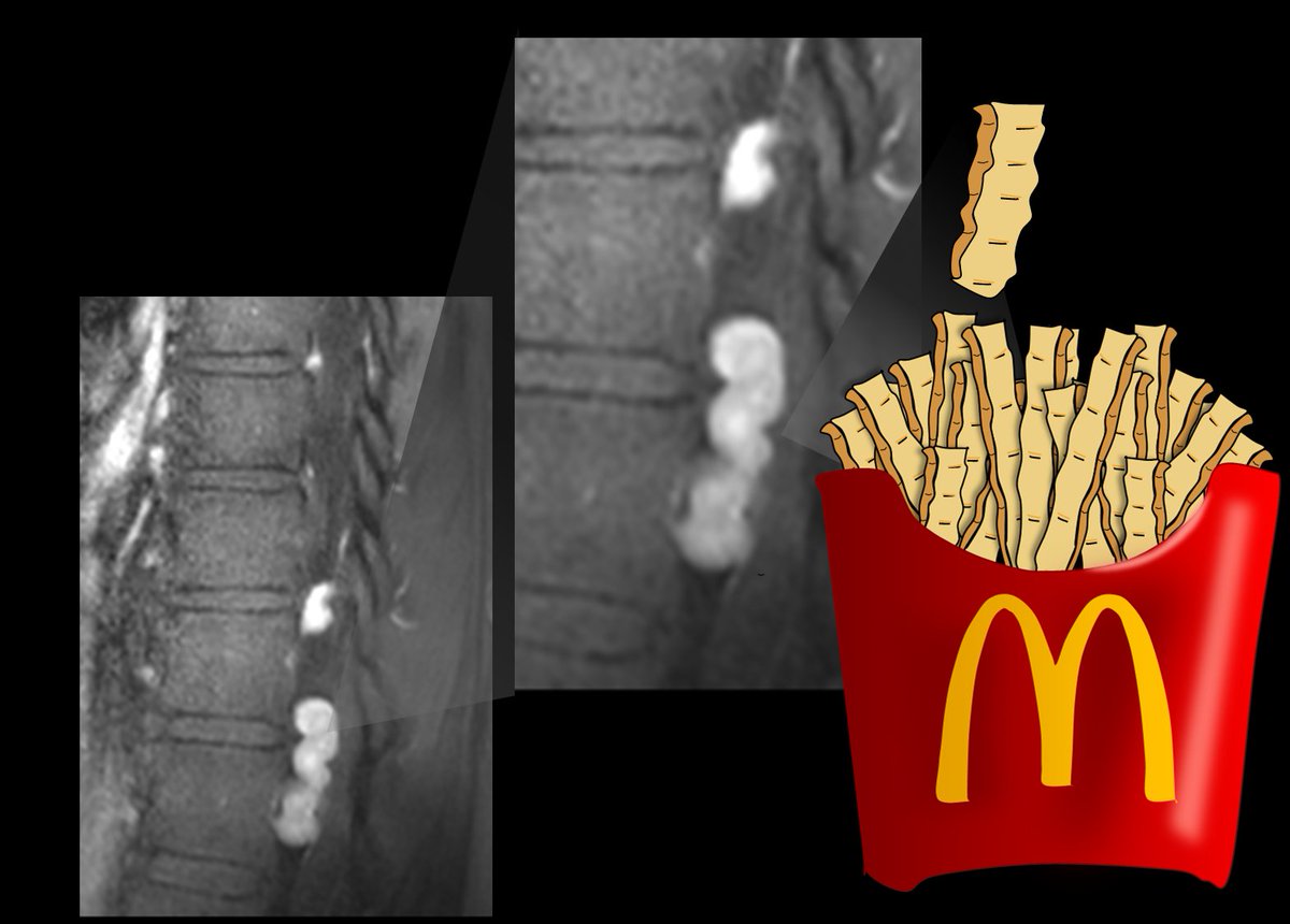
3/On the T2 weighted images, the lesions showed a very distinct donut-like appearance, with a very dark, almost black rim and a bright center. Image
4/On contrast imaging, the donuts were connected as twisty, tubular structures—my fellow said they looked like crinkle fries! (and yes, I know McDonald’s doesn’t serve crinkle fries as illustrated, but it’s just an illustration—there aren’t fries in the spine either 😉) Image
5/Tubular sounds like a vessel! And these did empty out into the neural foramina. But the spinal angio was negative for a vascular lesion (not all images shown—normal studies don’t make for good twitter images) Image
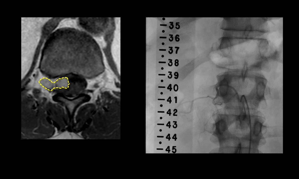
6/These are intradural varices! B/c of slow flow, they may not fill on angiography & lose their flow void centrally—causing a“donut sign.” Etiology is unclear, but likely acquired
So the next time you see donuts & fries in the spine—you can be sure you've got the right diagnosis Image

• • •

Missing some Tweet in this thread? You can try to force a refresh
 

Keep Current with Lea Alhilali, MD

Lea Alhilali, MD Profile picture

Stay in touch and get notified when new unrolls are available from this author!

Read all threads

This Thread may be Removed Anytime!

PDF

Twitter may remove this content at anytime! Save it as PDF for later use!

Try unrolling a thread yourself!

how to unroll video
  1. Follow @ThreadReaderApp to mention us!

  2. From a Twitter thread mention us with a keyword "unroll"
@threadreaderapp unroll

Practice here first or read more on our help page!

More from @teachplaygrub

Apr 19
1/Having trouble remembering how to differentiate dementias on imaging?

Is looking at dementia PET scans one of your PET peeves?

Here’s a thread to show you how to remember the imaging findings in dementia & never forget! Image
2/The most common functional imaging used in dementia is FDG PET. And the most common dementia is Alzheimer’s disease (AD).

On PET, AD demonstrates a typical Nike swoosh pattern—with decreased metabolism in the parietal & temporal regions Image
3/The swoosh rapidly tapers anteriorly—& so does hypometabolism in AD in the temporal lobe. It usually spares the anterior temporal poles.

So in AD look for a rapidly tapering Nike swoosh, w/hypometabolism in the parietal/temporal regions—sparing the anterior temporal pole Image
Read 16 tweets
Apr 18
1/”That’s a ninja turtle looking at me!” I exclaimed.

My fellow rolled his eyes, “Why do I feel I’m going to see this on X or twitter soon…”

He was right!

A thread about one of my favorite imaging findings & pathology behind it ! Image
2/Now the ninja turtle isn’t an actual sign—yet!

But I am hoping to make it go viral as one.

To understand what this ninja turtle is, you first have to know the anatomy in this region.

I have always thought the medulla looks like a 3 leaf clover in this region. Image
3/ The most medial bump of the clover is the medullary pyramid (motor fibers).

Next to it is the inferior olivary nucleus (ION), & finally, the last largest leaf is the inferior cerebellar peduncle.

Now you can see that the ninja turtle eyes correspond to the ION. Image
Read 11 tweets
Apr 17
1/CSF leaks are controversial!

Some say they're overdiagnosed, others underdiagnosed

How can YOU make sure you aren’t under or overdiagnosing?

Are you BERN-ing to know when to suspect CSF leak?

Here’s a 🧵about the CSF leak Bern score so you don’t get BERN-ed by CSF leaks Image
2/In CSF leaks, everyone knows about brain sagging.

But this can happen w/other diseases, ie Chiari 1.

Other findings can be seen on brain MRI in CSF leaks.

But what are these findings & are some findings more suggestive than others?

Do⬆️findings = ⬆️suspicion? Image
3/The Bern group looked at 9 quantitative & 7 qualitative signs seen on brain MRI in CSF leaks to see which are most important.

Depending on type & # of findings, they developed a score to indicate what level of suspicion you should have for a leak. Image
Read 15 tweets
Apr 15
1/Is remembering cerebellar anatomy making you dizzy?

Need help telling your flocculus from your nodule?

How much cerebellar anatomy do YOU know?

Here’s some help w/an anatomy thread on the 9 lobules of the vermis! Image
2/Coming from anterior, the first lobule is the lingula

It sticks out from the front of the vermis & is connected to the superior cerebellar peduncle (SCP)

I remember this bc of its very appropriate name—lingula—it looks like a tongue sticking out of the vermis to lick the SCP Image
3/Moving clockwise, next is the central lobule

I remember this bc it's positioned exactly how a central lobule should be positioned, in the driver’s seat!

It's where the front driver position would be if the vermis was a car—up front, looking out a windshield over the lingula Image
Read 12 tweets
Apr 12
1/Time is brain! But what time is it?

If you don’t know the time of stroke onset, are you able to deduce it from imaging?

Here’s a thread to help you date a stroke on MRI! Image
2/In ~25% of acute stroke patients, the time of last known well is well, not known.

Then it’s important to use the stroke’s MR imaging features to help date its timing.

Is it hyperacute? Acute? Subacute? Or are the “stroke” symptoms from a seizure from their chronic infarct? Image
3/Strokes evolve, or grow old, the same way people evolve or grow old.

Appearance of stroke on imaging mirrors the life stages of a person—you just have to change days for a stroke into years for a person.

So 15 day old stroke has features of a 15 year old person, etc. Image
Read 22 tweets
Apr 1
1/Does your ability to remember temporal lobe anatomy seem, well, temporary?

Or are you feeling temporally challenged when it comes to this complex region?

Here’s a thread to help you remember the structures of the temporal lobe! Image
2/Temporal lobe can be divided centrally & peripherally.

Centrally is the hippocampus.

It’s a very old part of the brain & is relatively well preserved going all the way back to rats.

Its main function is memory—getting both rats & us through mazes—including the maze of life Image
3/Peripherally is the neocortex.

Although rats also have neocortex, theirs is much different structurally than humans.

So I like to think of neocortex as providing the newer (neo) functions of the temporal lobes seen in humans: speech, language, visual processing/social cues Image
Read 12 tweets

Did Thread Reader help you today?

Support us! We are indie developers!


This site is made by just two indie developers on a laptop doing marketing, support and development! Read more about the story.

Become a Premium Member ($3/month or $30/year) and get exclusive features!

Become Premium

Don't want to be a Premium member but still want to support us?

Make a small donation by buying us coffee ($5) or help with server cost ($10)

Donate via Paypal

Or Donate anonymously using crypto!

Ethereum

0xfe58350B80634f60Fa6Dc149a72b4DFbc17D341E copy

Bitcoin

3ATGMxNzCUFzxpMCHL5sWSt4DVtS8UqXpi copy

Thank you for your support!

Follow Us!

:(