Lea Alhilali, MD Profile picture
Jun 17, 2022 20 tweets 10 min read Read on X
1/Radiologist not answering the phone?Just want a quick read on that stat head CT?

Here's a little help on how to do it yourself w/a #tweetorial on how to read a head CT!
#medtwitter #FOAMed #FOAMrad #medstudenttwitter #medstudent #neurorad #radres @MedTweetorials #neurosurgery Image
2/In bread & butter neuroimaging—CT is the bread—maybe a little bland, not super exciting—but necessary & you can get a lot of nutrition out of it. MRI is like the butter—everyone loves it, it makes everything better, & it packs a lot of calories. Today, we start w/the bread! Image
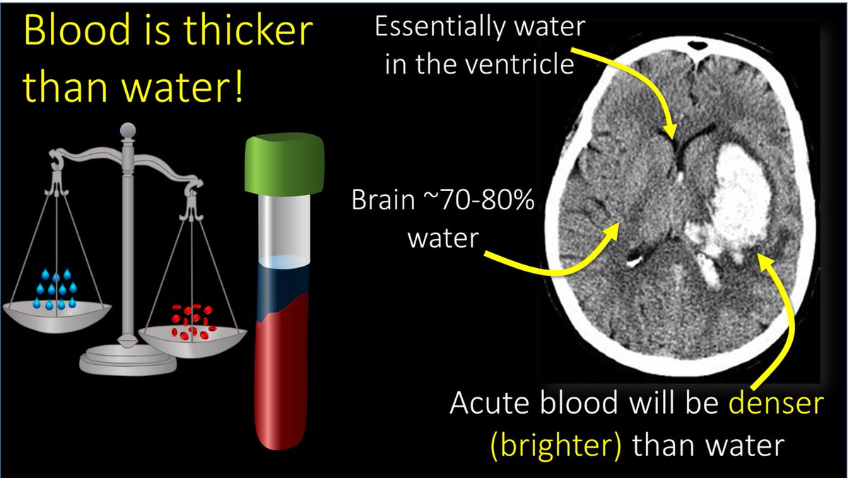
3/The most important thing to look for on a head CT is blood. Blood is Bright on a head CT—both start w/B. Blood is bright bc for all it’s Nobel prizes, all CT is is a density measurement—and blood is denser (thicker) than water and denser things are brighter on CT Image
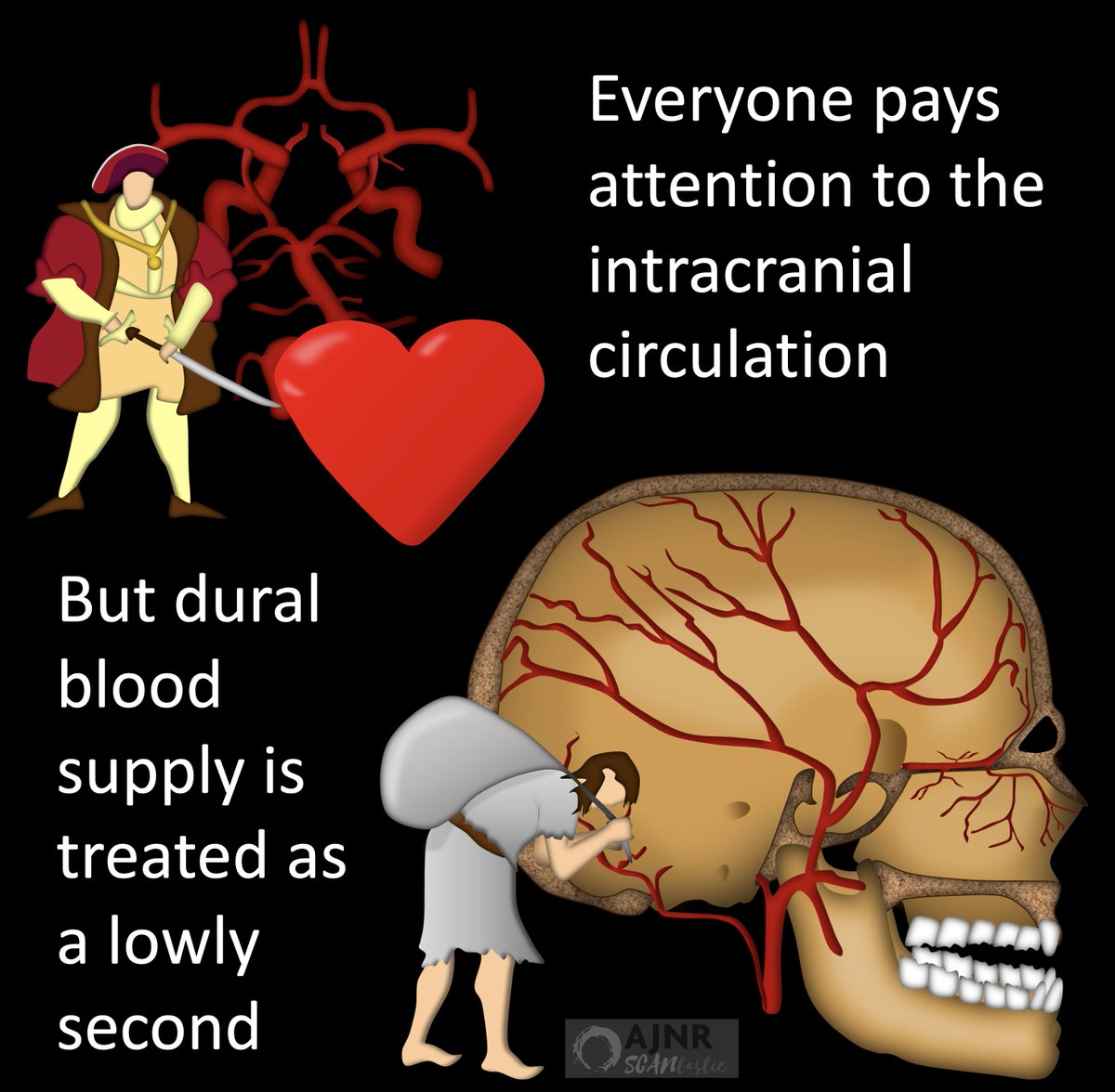
4/Once you see blood, the next question is—where is it? To know this, we need to know meningeal layers. Outer most layer is the dura mater. I remember it bc dura mater is DURAble. It is thick like a winter coat. Like a winter coat, it doesn’t hug the curves & hides rolls of fat. Image
5/Inner most layer is the pia mater. It is thin and hugs the curves of the brain like an adult onsie. I remember it bc pee-ah mater is just a few letters away from pee-jay mater—so it sounds like adult onsie PJs Image
6/In between these two layers is the arachnoid. It is called that because it contains web like septations like a spider’s web (ARACHnoid like ARACHnophobia). So now you know the meningeal layers. I remember the order bc the meninges “P-A-D” the brain—Pia/Arachnoid/Dura Image
7/Blood can be anywhere in these layers. EPIdural is beside the dura, or outside all layers. SUBdural is below the dura, but still outside pia & arachnoid. SUBarachnoid is below both dura & arachnoid. I’m skipping intraparenchymal hemorrhage here bc that is relatively obvious. Image
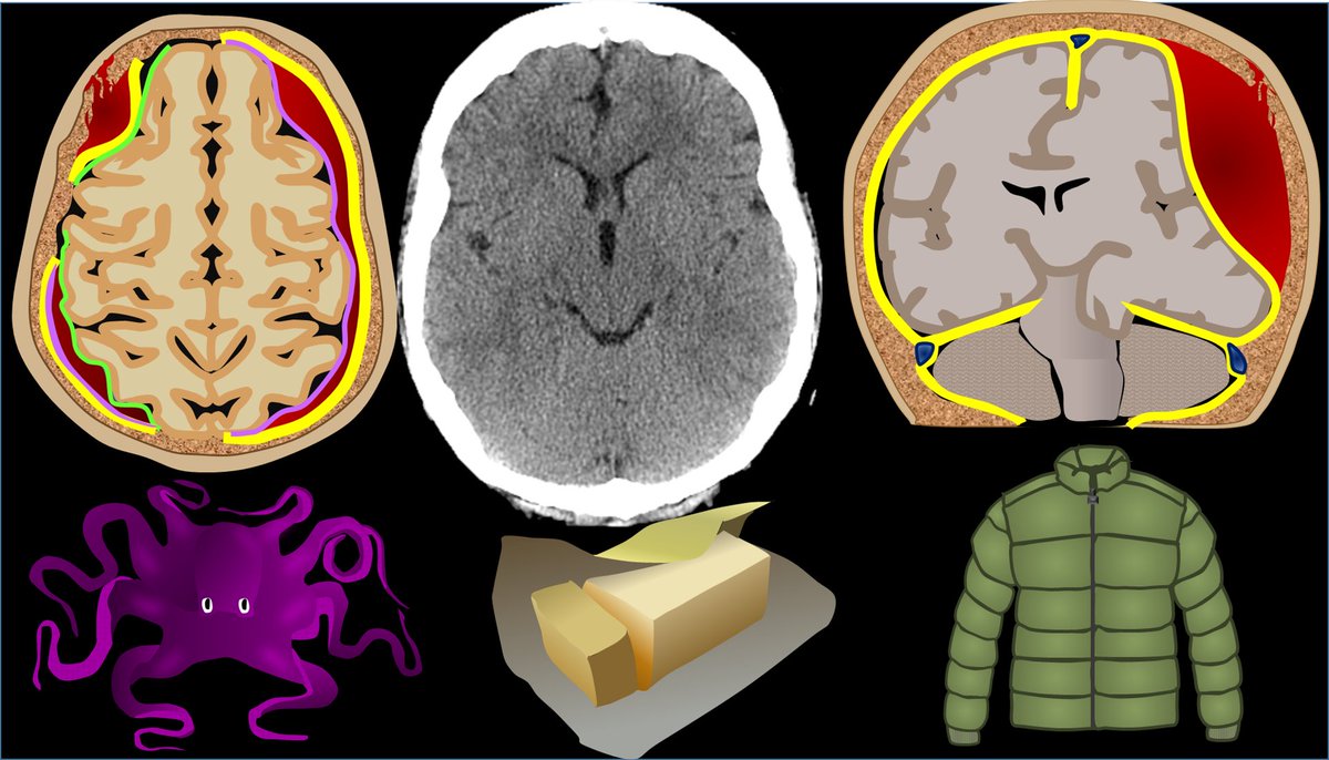
8/Each of these types of hemorrhage has a unique look on CT. Epidural hemorrhage is called “lentiform” bc it is convex out like a lens or a pregnant belly. Subdural hemorrhage wraps around the brain like a crescent. Subarachnoid hemorrhage is curvy between gyri like a snake Image
9/So why is intracranial hemorrhage so dangerous? You won’t exsanguinate from intracranial hemorrhage like a retroperitoneal bleed. The reason intracranial hemorrhage is so dangerous is bc the calvarium is a closed space with no give for anything extra. Image
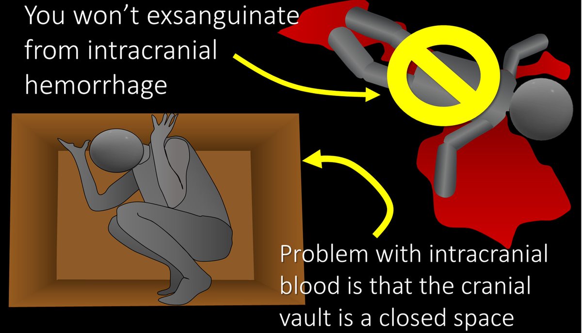
10/So when you add something extra like blood, the calvarium won’t give, and something else has to—and that’s the brain. Blood will push on the brain causing damage from the associated mass effect. Image
11/Let’s talk about mass effect. Symmetry is beautiful—that’s why Denzel Washington is such the epitome of beauty bc he is perfectly symmetry. The brain on a CT should be symmetric. A CT tech once told me he could make all the findings on CTs bc all he did was look for asymmetry. Image
12/So on every CT you should look for symmetry—and things that are asymmetric are BAD. If you can’t draw a line down the middle have each side be a mirror image—something is wrong. Image
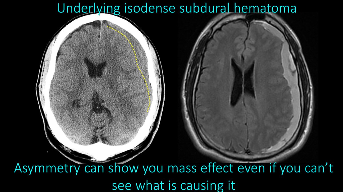
13/This asymmetry was from an subdural hemorrhage that was the same density as brain—making it difficult to visualize, but you could tell it was there from the asymmetry it caused. Mass effect causes asymmetry Image
14/Mass effect can cause brain to herniate into wrong compartments. There are 2 main herniation types. Subfalcine herniation is where one side slides under the falx to the other side. On CT, we call this midline shift—how much one side shifts under the midline to the other side Image
15/Next is transtentorial herniation—where the supratentorial compartment herniates through the tentorium that separates the cerebral hemispheres from the cerebellum. We see this on CT by effacement of the basilar cisterns—which are CSF spaces at the base of the brain. Image
16/The two most important cisterns for herniation are the suprasellar cistern—which looks like a pentagon—and the ambient/quadrigeminal cistern that look like the mouth of a semi-evil smiley face with the lateral and third ventricles as the eyes and nose. Image
17/With transtentorial herniation, we are looking for that pentagon to become a triangle or that smiley to get a Bell’s palsy—with part of it missing. If you see either of those, there is transtentorial herniation. Image
18/The final thing to look for on a head CT is a stroke. We see this as loss of gray-white differentiation. Normally, the interface between gray and white matter is crisp and looks like long octopus arms of white matter reaching out into the gray matter. Image
19/With a stroke, this interface gets blurred. It is like some took a painting that had a clear line between the white and gray matter and just smeared the white matter into the gray matter. If I see anywhere where the white matter looks smeared into the gray, I call an infarct Image
20/So now you know the basics of head CTs! Hopefully now your reads of the bread of neuroimaging will go smoothly like butter! Image

• • •

Missing some Tweet in this thread? You can try to force a refresh
 

Keep Current with Lea Alhilali, MD

Lea Alhilali, MD Profile picture

Stay in touch and get notified when new unrolls are available from this author!

Read all threads

This Thread may be Removed Anytime!

PDF

Twitter may remove this content at anytime! Save it as PDF for later use!

Try unrolling a thread yourself!

how to unroll video
  1. Follow @ThreadReaderApp to mention us!

  2. From a Twitter thread mention us with a keyword "unroll"
@threadreaderapp unroll

Practice here first or read more on our help page!

More from @teachplaygrub

Jul 2
1/The medulla is anything but DULL!

Does seeing an infarct in the medulla cause your heart to skip a beat?

Does medullary anatomy send you into respiratory arrest?

Never fear, here is a thread on the major medullary syndromes! Image
2/The medulla is like a toll road.

Everything going down into the cord must pass through the medulla & everything from the cord going back up to the brain must too.

That’s a lot of tracts for a very small territory. Luckily you don’t need to know every tract Image
3/Medulla has 4 main vascular territories, spread out like a fan: anteromedial, anterolateral, lateral, and posterior.

You don’t need to remember their names, just the territory they cover—and I’ll show you how Image
Read 18 tweets
Jun 30
1/Time is brain! But what time is it?

If you don’t know the time of stroke onset, are you able to deduce it from imaging?

Here’s a thread to help you date a stroke on MRI! Image
2/Strokes evolve, or grow old, the same way people evolve or grow old.

The appearance of stroke on imaging mirrors the life stages of a person—you just have to change days for a stroke into years for a person

So 15 day old stroke has features of a 15 year old person, etc. Image
3/Initially (less than 4-6 hrs), the only finding is restriction (brightness) on diffusion imaging (DWI).

You can remember this bc in the first few months, a baby does nothing but be swaddled or restricted. So early/newly born stroke is like a baby, only restricted Image
Read 10 tweets
Jun 27
1/”I LOVE spinal cord syndromes!” is a phrase that has NEVER, EVER been said by anyone.

Do you become paralyzed when you see cord signal abnormality?

Never fear—here is a thread on all the incomplete spinal cord syndromes to get you moving again! Image
2/Spinal cord anatomy can be complex. On imaging, we can see the ant & post nerve roots. We can also see the gray & white matter. Hidden w/in the white matter, however, are numerous efferent & afferent tracts—enough to make your head spin. Image
3/Lucky for you, for the incomplete cord syndromes, all you need to know is gray matter & 3 main tracts. Anterolaterally, spinothalamic tract (pain & temp). Posteriorly, dorsal columns (vibration, proprioception, & light touch), & next to it, corticospinal tracts—providing motor Image
Read 20 tweets
Jun 23
1/Do you get a Broca’s aphasia trying remember the location of Broca's area?

Does trying to remember inferior frontal gyrus anatomy leave you speechless?

Don't be at a loss for words when it comes to Broca's area

Here’s a 🧵to help you remember the anatomy of this key region! Image
2/Anatomy of the inferior frontal gyrus (IFG) is best seen on the sagittal images, where it looks like the McDonald’s arches.

So, to find this area on MR, I open the sagittal images & scroll until I see the arches. When it comes to this method of finding the IFG, i’m lovin it. Image
3/Inferior frontal gyrus also looks like a sideways 3, if you prefer. This 3 is helpful bc the inferior frontal gyrus has 3 parts—called pars Image
Read 13 tweets
Jun 19
1/Feeling intoxicated trying to remember all the findings in alcohol use disorder?!

Here’s something to put you in high spirits!

This month’s @Radiographics has the important neuroimaging findings alcohol use disorder!


@cookyscan1 @RadG_editor #RGphx pubs.rsna.org/doi/10.1148/rg…Image
2/There’s an easy rhyme to help you remember the important neuroimaging findings of alcohol use disorder

“Basal ganglia is white...”

Get intrinsic T1 shortening in the BG that makes it look white as a ghost! Image
3/Next “...Cortex is bright”

Acute hyperammonemic encephalopathy cause cortical restricted diffusion, especially the insula, so that it looks as bright as a light bulb! Image
Read 8 tweets
Jun 9
1/Need help reading spine imaging? I’ve got your back!

It’s as easy as ABC!

A thread about an easy mnemonic you can use on every single spine study you see to increase your speed & make sure you never miss a thing! Image
2/A is for alignment

Look for:
(1) Unstable injuries

(2) Malalignment that causes early degenerative change. Abnormal motion causes spinal elements to abnormally move against each other, like grinding teeth wears down teeth—this wears down the spine Image
3/B is for bones.

On CT, the most important thing to look for w/bones is fractures. You may see focal bony lesions, but you may not

On MR, it is the opposite—you can see marrow lesions easily but you may or may not see edema associated w/fractures if the fracture is subtle Image
Read 11 tweets

Did Thread Reader help you today?

Support us! We are indie developers!


This site is made by just two indie developers on a laptop doing marketing, support and development! Read more about the story.

Become a Premium Member ($3/month or $30/year) and get exclusive features!

Become Premium

Don't want to be a Premium member but still want to support us?

Make a small donation by buying us coffee ($5) or help with server cost ($10)

Donate via Paypal

Or Donate anonymously using crypto!

Ethereum

0xfe58350B80634f60Fa6Dc149a72b4DFbc17D341E copy

Bitcoin

3ATGMxNzCUFzxpMCHL5sWSt4DVtS8UqXpi copy

Thank you for your support!

Follow Us!

:(