Lea Alhilali, MD Profile picture
Jun 27, 2022 22 tweets 10 min read Read on X
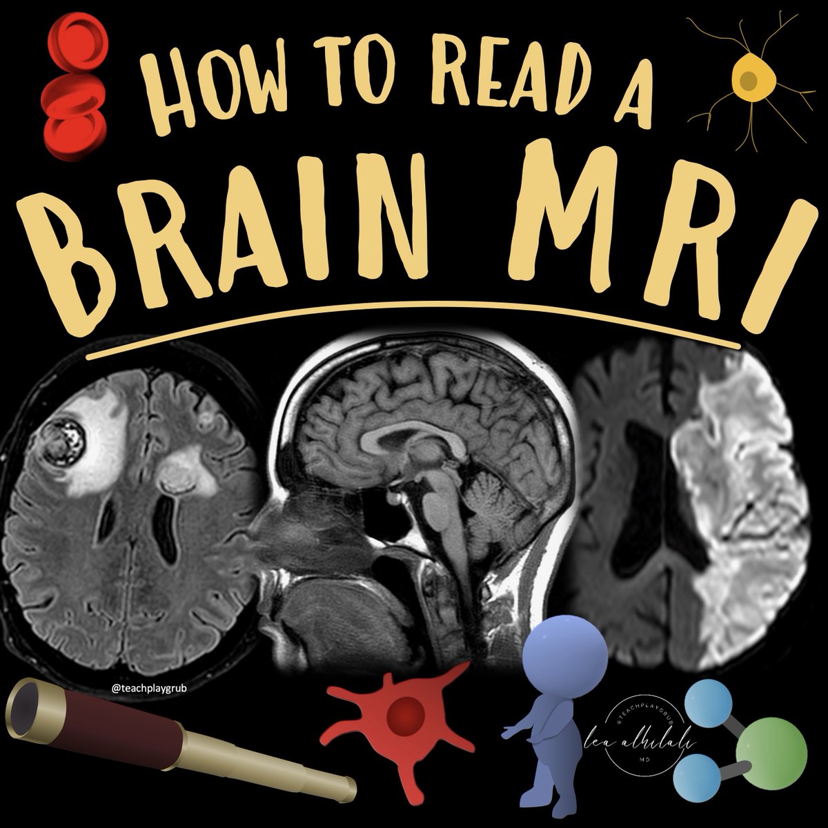
1/Do radiologists sound like they are speaking a different language when they talk about MRI? T1 shortening what?T2 prolongation who?
Here’s a translation w/a #tweetorial introduction to MRI
#medtwitter #FOAMed #FOAMrad #medstudent #neurorad #radres @MedTweetorials #neurosurgery Image
2/When it comes to bread and butter neuroimaging—MRI is definitely the butter. Butter makes everything taste better and packs a lot of calories. MRI can add so much information to a case Image
3/In fact, if CT is a looking glass into the brain—MRI is a microscope. It can tell us so much more about the brain and pathology that affects the brain. So let’s talk about the basic sequences that make up an MRI and what they can show us. Image
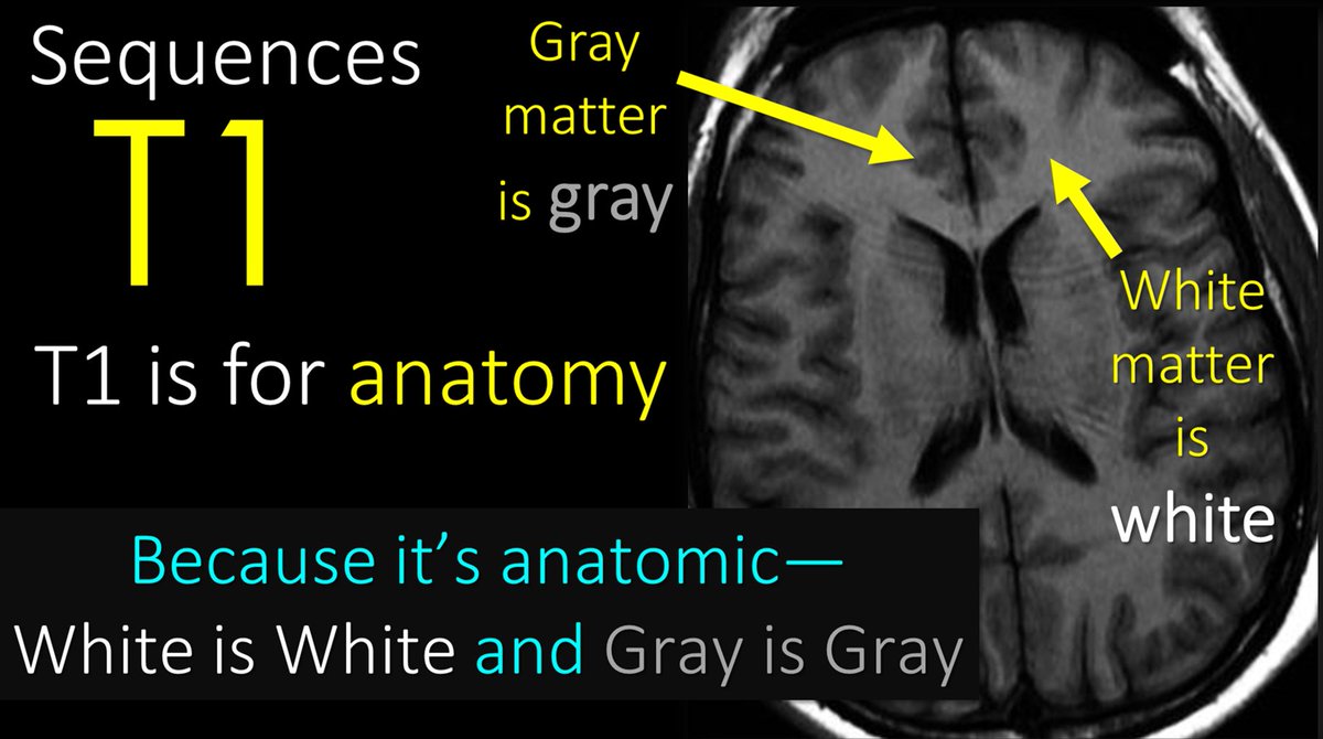
4/Let’s start w/T1—it is #1 after all! T1 is for anatomy. Since it’s anatomic, brain structures will reflect the same color as real life. So gray matter is gray on T1 & white matter is white on T1. So if you see an image where gray is gray & white is white—you know it’s a T1. Image
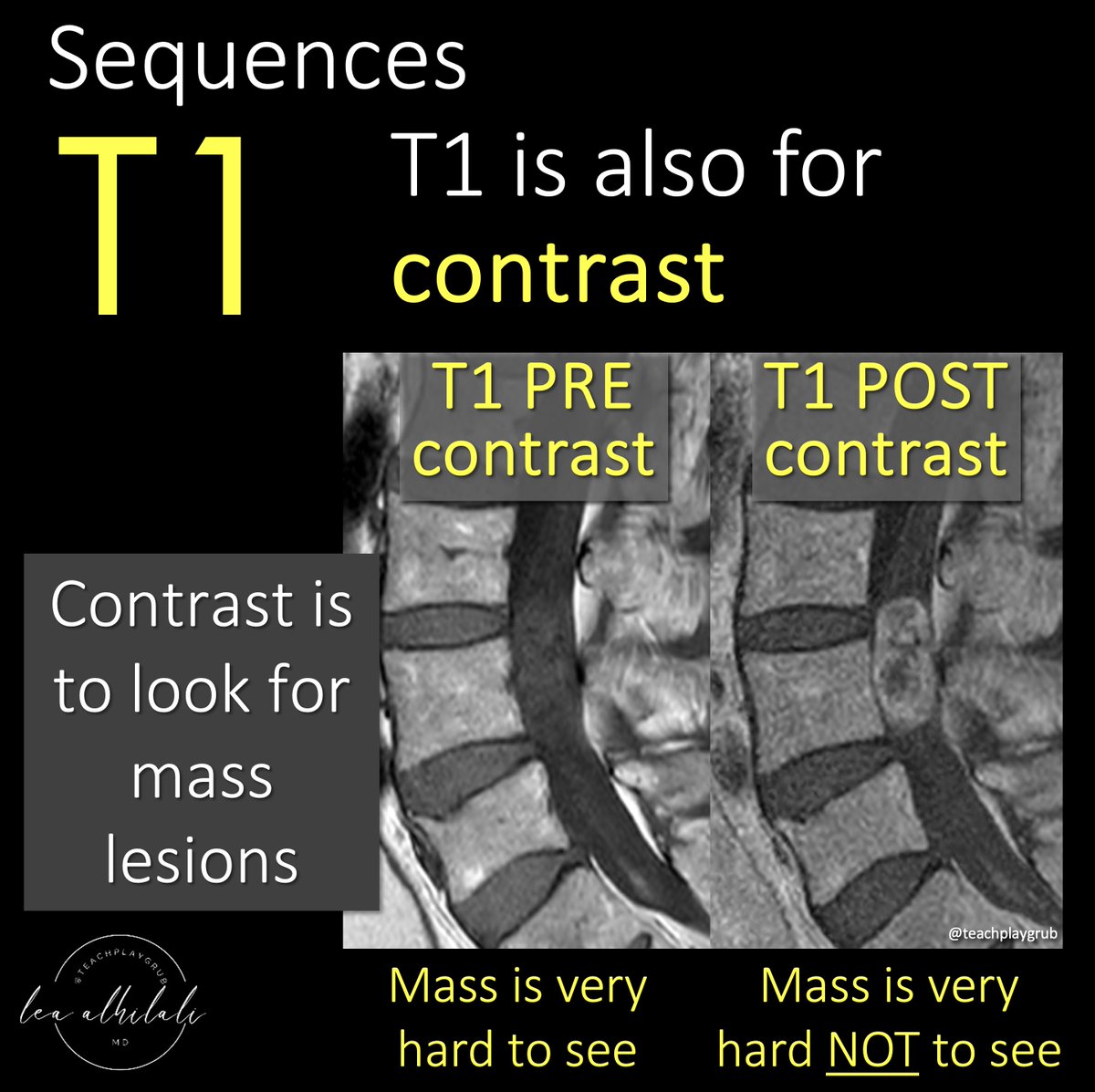
5/T1 is also for contrast. Contrast material helps us to see masses. Contrast can’t get into normal brain & spine bc of the blood brain barrier—but masses don’t have a blood brain barrier, so when you give contrast, masses will take it up & light up, making them easier to see. Image
6/So to review, T1 is for anatomy and contrast. I remember this bc anatomy is the number 1 thing a radiologist needs to know and a mass is the number 1 thing a radiologist doesn’t want to miss. Image
7/Now to T2! T2 sequences are water sensitive sequences. What is pathologic water in the brain? Edema! My attending once said, “Everything bad in this world is trying to turn you back into what you came from—water. So T2 shows you edema—but this edema can be from many things. Image
8/To review—T1 is for anatomy and contrast, T2 (and FLAIR, which is a type of T2) is for water—which is bright on T2. I remember this bc H20 has a 2 in it—T2 is for H20. Image
9/Next to diffusion or DWI. Diffusion is primarily to detect stroke. Acute strokes are bright on diffusion. But just as all that glitters is not gold, not all that is bright on DWI is an acute stroke. Image
10/This is bc all diffusion does is detect how difficult it is for water to move. Anything that makes the space around water crowded and difficult to move will be bright on diffusion imaging Image
11/So classically, it from a stroke. When cells run out of ATP, the Na/K pump stops working & immediately water rushes in from osmotic pressure & the cells swell. These swollen cells fill the interstitium & restrict the movement of water. This is why strokes are bright on DWI! Image
12/But other things can make it crowded and difficult for water to move. For example, tightly packed cells in aggressive tumors will also fill the spaces & make it difficult for water to move—it is trapped between the tumor cells! So highly cellular tumors are often bright on DWI Image
13/Here is an example. Here is a mass that is as bright as stroke on diffusion bc of its densely packed cells. On contrast images, we see it avidly enhance, as we would expect for a mass. On CT, the tumor is very dense bc of the densely packed cells. Image
14/Hematomas are also bright on DWI. In normal blood, water flows happy & free—but once the clotting cascade starts & fibrin & thrombin & whatever stuff I don’t remember as a radiologist clumps everything together, things get tight—water is trapped in the clot interstices! Image
15/Here is an example. The hemorrhage is bright on CT bc it is clotted, and thus more dense than the brain and CSF, which are closer in density to water. For this same reason, the hemorrhage is bright on diffusion—bc the dense clot traps the water. Image
16/Pus is also bright on diffusion. As a radiologist I don’t often see pus, but as a mom, I sure do. It is thick and gooey and you can just imagine how difficult it is for water to travel through that gelantinous blob of pus. Image
17/Here’s an example. There is a ring enhancing lesion w/a lot of edema on T2. Centrally, there is restricted diffusion, meaning that there is something gooey or thick or dense centrally. Bc this central stuff doesn’t enhance, we know it’s not a mass. This is pus in an abscess! Image
18/So to review--while not everything that is bright on diffusion is a stroke, the most important use is for strokes. I remember this bc it's called DWI--which I joking say stands for Diagnose With Infarct! Image
19/Last but not least is gradient imaging. Gradient imaging is sensitive to metals. And what’s the most important metal in body? Iron—bc iron is in blood. So gradient is our blood sensitive sequence Image
20/Blood is black on gradient. I remember this bc gradient is for metal—and when I think of metal, I think of blacksmiths forging metal products. So BLACKsmith=metal is BLACK on gradient. Image
21/But other metals will be black too. Notably, calcium, which is in our bones and in many other lesions. So remember, just all that glitters is not gold, not all that is black on gradient is blood—other metals are black too Image
22/So now you know the basic MRI sequences and what they are used for. So hopefully now, the radiologist won’t sound like they are speaking a different language when they talk to you—they will just be nerdy and socially awkward when they do! Image

• • •

Missing some Tweet in this thread? You can try to force a refresh

 

Keep Current with Lea Alhilali, MD

Lea Alhilali, MD Profile picture

Stay in touch and get notified when new unrolls are available from this author!

Read all threads

This Thread may be Removed Anytime!

PDF

Twitter may remove this content at anytime! Save it as PDF for later use!

Try unrolling a thread yourself!

how to unroll video
  1. Follow @ThreadReaderApp to mention us!

  2. From a Twitter thread mention us with a keyword "unroll"
@threadreaderapp unroll

Practice here first or read more on our help page!

More from @teachplaygrub

Jun 29
1/I always say you can tell a bad read on a spine MR if it doesn’t talk about lateral recesses.

What will I think when I see your read? Do you rate lateral recess stenosis?

Here’s a thread on lateral recess anatomy & a grading system for lateral recess stenosis Image
2/First anatomy.

Thecal sac is like a highway, carrying the nerve roots down the lumbar spine.

Lateral recess is part of the lateral lumbar canal, which is essentially the exit for spinal nerve roots to get off the thecal sac highway & head out into the rest of the body Image
3/Exits have 3 main parts.

First is the deceleration lane, where the car slows down as it starts the process of exiting.

Then there is the off ramp itself, and this leads into the service road which takes the car to the roads that it needs to get to its destination Image
Read 21 tweets
Jun 26
1/Time is brain! But what time is it?

If you don’t know the time of stroke onset, are you able to deduce it from imaging?

Here’s a thread to help you date a stroke on MRI! Image
2/Strokes evolve, or grow old, the same way people evolve or grow old

The appearance of stroke on imaging mirrors the life stages of a person—you just have to change days for a stroke into years for a person

So 15 day old stroke has features of a 15 year old person, etc. Image
3/Initially (less than 4-6 hrs), the only finding is restriction (brightness) on diffusion imaging (DWI)

You can remember this bc in the first few months, a baby does nothing but be swaddled or restricted

So early/newly born stroke is like a baby, only restricted Image
Read 10 tweets
Jun 1
1/Having trouble remembering how to differentiate dementias on imaging?

Is looking at dementia PET scans one of your PET peeves?

Here’s a thread to show you how to remember the imaging findings in dementia & never forget! Image
2/The most common functional imaging used in dementia is FDG PET. And the most common dementia is Alzheimer’s disease (AD).

On PET, AD demonstrates a typical Nike swoosh pattern—with decreased metabolism in the parietal & temporal regions Image
3/The swoosh rapidly tapers anteriorly—& so does hypometabolism in AD in the temporal lobe. It usually spares the anterior temporal poles.

So in AD look for a rapidly tapering Nike swoosh, w/hypometabolism in the parietal/temporal regions—sparing the anterior temporal pole Image
Read 16 tweets
May 1
1/Do radiologists sound like they are speaking a different language when they talk about MRI?

T1 shortening what? T2 prolongation who?

Here’s a translation w/an introductory thread to MRI. Image
2/Let’s start w/T1—it is #1 after all! T1 is for anatomy

Since it’s anatomic, brain structures will reflect the same color as real life

So gray matter is gray on T1 & white matter is white on T1

So if you see an image where gray is gray & white is white—you know it’s a T1 Image
3/T1 is also for contrast

Contrast material helps us to see masses

Contrast can’t get into normal brain & spine bc of the blood brain barrier—but masses don’t have a blood brain barrier, so when you give contrast, masses will take it up & light up, making them easier to see. Image
Read 20 tweets
Apr 30
1/Does your ability to remember temporal lobe anatomy seem, well, temporary?

Or are you feeling temporally challenged when it comes to this complex region?

Here’s a thread to help you remember the structures of the temporal lobe! Image
2/Temporal lobe can be divided centrally & peripherally.

Centrally is the hippocampus.

It’s a very old part of the brain & is relatively well preserved going all the way back to rats.

Its main function is memory—getting both rats & us through mazes—including the maze of life Image
3/Peripherally is the neocortex.

Although rats also have neocortex, theirs is much different structurally than humans.

So I like to think of neocortex as providing the newer (neo) functions of the temporal lobes seen in humans: speech, language, visual processing/social cues Image
Read 12 tweets
Mar 25
1/How low can you go??

All the hype nowadays is about high field MRI, but what about low field??

Read on for this month’s @theAJNR SCANtastic for what to know about what may be the next biggest thing in MRI!

ajnr.org/content/47/3/7…Image
2/The growing strength is for larger & larger field strengths for higher & higher resolution

So why would we possible go backwards to lower field strength?

Turns out there are some advantages. Image
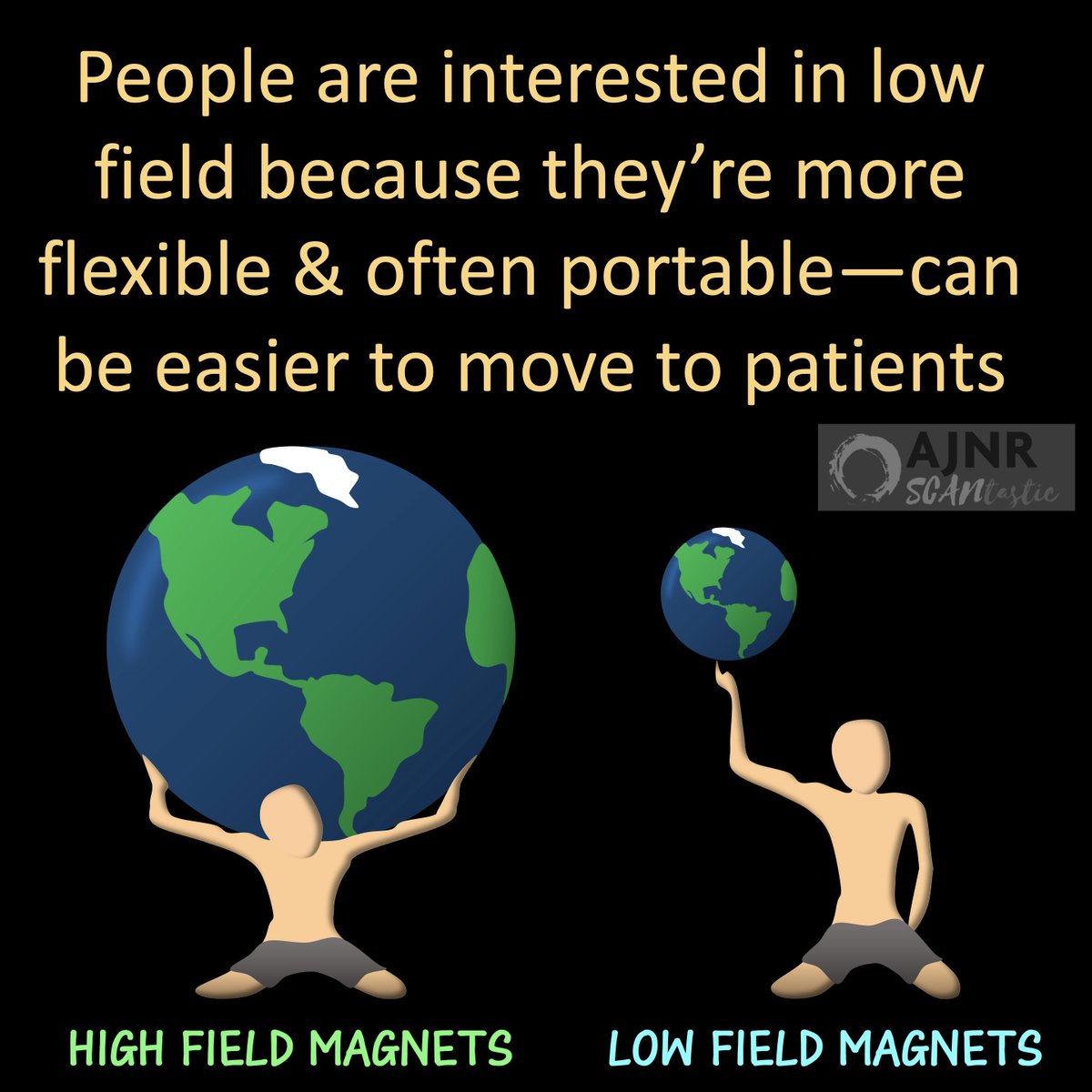
3/Low field strength magnets are much for flexible

They can be put in non-traditional settings (clinics) & can also possibly be moved to the bedside

It is truly POC MRI!

But how does it perform? Image
Read 11 tweets

Did Thread Reader help you today?

Support us! We are indie developers!


This site is made by just two indie developers on a laptop doing marketing, support and development! Read more about the story.

Become a Premium Member ($3/month or $30/year) and get exclusive features!

Become Premium

Don't want to be a Premium member but still want to support us?

Make a small donation by buying us coffee ($5) or help with server cost ($10)

Donate via Paypal

Or Donate anonymously using crypto!

Ethereum

0xfe58350B80634f60Fa6Dc149a72b4DFbc17D341E copy

Bitcoin

3ATGMxNzCUFzxpMCHL5sWSt4DVtS8UqXpi copy

Thank you for your support!

Follow Us!

:(