Lea Alhilali, MD Profile picture
Jul 22, 2022 10 tweets 6 min read Read on X
1/Remembering spinal fracture classifications is back breaking work!

A #tweetorial to review the scoring system for thoracic & lumbar fractures—“TLICS” to the cool kids! #medstudenttwitter #medtwitter #radres #FOAMed #FOAMrad #neurorad #Meded #backpain #spine #Neurosurgery
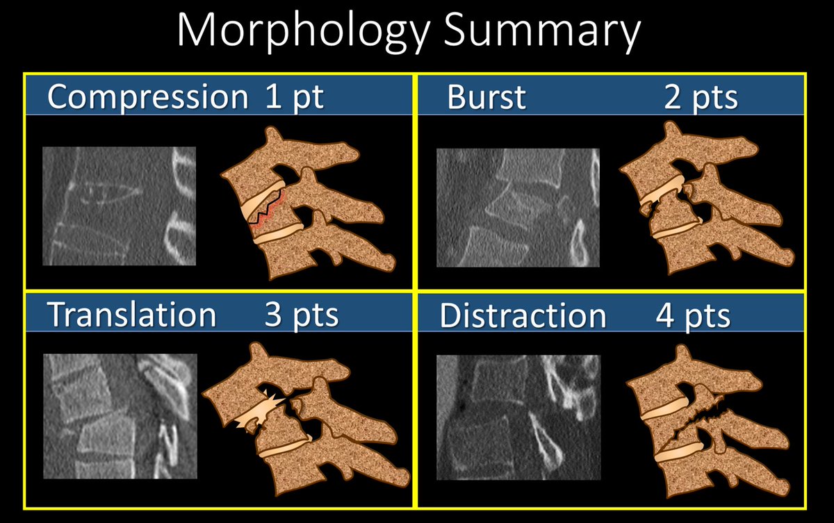
2/We’ll talk about the imaging part of TLICS. TLICS scores a fx on (1) morphology & (2) posterior ligamentous complex (PLC) injury. Let’s start w/morphology. W/only mild axial loading, you get the simplest fx, a compression fx—like a simple long bone fx--worth 1 pt.
3/As the axial force grows, this becomes a burst fx with retropulsion of the posterior vertebral body—just as greater force causes more comminution in long bone fxs. A burst is worth 2 points.
4/If the force is shearing, rather simply compress a vertebral body, you rip the connection between the vertebral bodies—this is the equivalent of pulling on a long bone & causing its dislocation from its joint or connection with another bone. This is worth 3 points
5/Similar to shear forces, distracting forces will rip the vertebral bodies apart. But rather than sliding forward or back, the vertebral bodies are pulled up or down, resulting in a vertical gap between the vertebral bodies. This is worth 4 points
6/This summarizes the TLICS scoring for fracture morphology. The higher the number, the greater the force and injury to the spine—ranging from simple compression fxs worth only 1 point to where the spine is literally ripped apart—a distraction injury, worth 4 points.
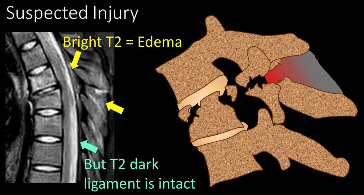
7/The next TLICS imaging finding is the integrity of the PLC. If it is intact, you get 0 pts. If you needed a tweetorial for that, well…I can’t help you much. If there is edema, but no true rupture on MRI, that is worth 2 pts. True disruption on MRI is worth 3 pts.
8/Here is an example of suspected injury—edema is seen in the posterior ligamentous complex, but the T2 dark lines that are the ligaments themselves appear intact. This is worth 2 pts.
9/If you can find a true disruption or gap in the T2 dark line of the ligament, that is considered truly disrupted and worth 3 points.
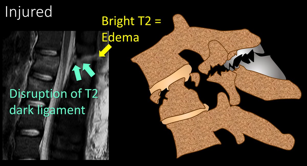
10/Here is the summary of the scoring for PLC injury in TLICS. Edema is suspicious, but only a true gap is considered injured.

So now you know how to score imaging findings in thoracolumbar fxs--I hope that takes a load off your back!

• • •

Missing some Tweet in this thread? You can try to force a refresh
 

Keep Current with Lea Alhilali, MD

Lea Alhilali, MD Profile picture

Stay in touch and get notified when new unrolls are available from this author!

Read all threads

This Thread may be Removed Anytime!

PDF

Twitter may remove this content at anytime! Save it as PDF for later use!

Try unrolling a thread yourself!

how to unroll video
  1. Follow @ThreadReaderApp to mention us!

  2. From a Twitter thread mention us with a keyword "unroll"
@threadreaderapp unroll

Practice here first or read more on our help page!

More from @teachplaygrub

Sep 15
1/Time is brain!

So you don’t have time to struggle w/that stroke alert head CT.

Here’s a thread to help you with the CT findings in acute stroke! Image
2/CT in acute stroke has 2 main purposes

(1) exclude hemorrhage (a contraindication to thrombolysis)

(2) exclude other pathologies mimicking acute stroke. But you can also see other findings to help diagnosis a stroke. Image
3/Infarct appearance depends on timing.

In first 12 hrs, the most common imaging finding is…a normal head CT

However, you may see a hyperdense artery or basal ganglia obscuration. Later, you see loss of gray white differentiation & sulcal effacement Image
Read 13 tweets
Sep 12
1/Do you feel there’s a back-log of findings in a spine MRI report?

Everyone talks about discs & facets, but not everyone talks about the endplates

Do you?

Do you need to talk about degenerative changes (Modic changes) of the endplates?

Here’s thread w/all you need to know! Image
2/Over 30 years ago, Modic et al. found there were 3 types of degenerative endplate changes:

(1) T2 bright changes (indicating edema, Modic 1)
(2) T1 bright changes (indicating fat, Modic 2)
(3) T1 & T2 dark changes (indicating sclerosis, Modic 3)

But what do they mean? Image
3/Let’s start w/Modic 1.

These are bright on T2, indicating edema

On pathology, it’s what you’d expect w/edema: inflammation, vascular granulation tissue, & high cellular turnover

Vascular granulation tissue means these can enhance on post contrast images—mimicking discitis! Image
Read 18 tweets
Sep 10
1/Are you FISHING for a way to better evaluate subarachnoid hemorrhage?

Are you hungry for a way to classify these patients?

Donut you worry!

Here’s a short thread to help you remember the modified Fisher scale for classifying subarachnoid hemorrhage. Image
2/Just think of the brain as a donut. Like a donut, it’s a bunch of stuff around a hole in the middle.

Ventricles are the hole in the middle of the brain just like there’s a hole in the middle of the dough in a donut.

Just don’t quote me to your neuroanatomy professor…. Image
3/Subarachnoid hemorrhage (SAH) added to the brain makes it less healthy, the same way adding toppings to a donut makes it less healthy.

Increasing severity of SAH is like increasingly unhealthy donut toppings. Fisher scale quantifies the vasospasm risk for increasing SAH Image
Read 8 tweets
Sep 8
1/Talk about twisting your back!

Do spine vascular lesions make your brain feel as tangled as the dilated vessels you see?

Want some more information on malformations?

Here’s a thread on spine vascular anatomy to give you durable knowledge on dural arteriovenous fistulas (dAVF)Image
2/To understand spinal dural AVFs, you need to understand basic spinal vascular anatomy.

The spine is LONG—to get blood from the top to the bottom is like going through the length of a marathon course Image
3/So we will need to tackle it like you tackle running a marathon.

When you run a marathon, you replenish yourself at aid/water stations along the way so you can make it all the way through.

Same w/spinal arterial vasculature—it needs to be replenished on the way down. Image
Read 19 tweets
Sep 3
1/Does the work up for dizziness make your head spin?

Wondering what to look for on an MR for dizziness

This month’s @theAJNR SCANtastic will tell you all you need about imaging Meniere’s disease!

ajnr.org/content/46/8/1…Image
@TheAJNR 2/The etiology for dizziness can have very diverse causes—each with very different treatments.

So it is important to try to differentiate

Meniere’s is a common cause & we can help diagnose it w/imaging! Image
@TheAJNR 3/To understand Meniere’s disease, you must know labyrinth anatomy

It has layers, like Russian nesting dolls. Outer doll is the bony labyrinth, holding perilymph & a second doll—membranous labyrinth.

Inside the membranous labyrinth is endolymph Image
Read 13 tweets
Aug 1
1/They say form follows function!

Brain MRI anatomy is best understood in terms of both form & function.

Here’s a short thread to help you to remember important functional brain anatomy--so you truly can clinically correlate! Image
2/Let’s start at the top. At the vertex is the superior frontal gyrus. This is easy to remember, bc it’s at the top—and being at the top is superior. It’s like the superior king at the top of the vertex. Image
3/It is also easy to recognize on imaging. It looks like a big thumb pointing straight up out of the brain. I always look for that thumbs up when I am looking for the superior frontal gyrus (SFG) Image
Read 12 tweets

Did Thread Reader help you today?

Support us! We are indie developers!


This site is made by just two indie developers on a laptop doing marketing, support and development! Read more about the story.

Become a Premium Member ($3/month or $30/year) and get exclusive features!

Become Premium

Don't want to be a Premium member but still want to support us?

Make a small donation by buying us coffee ($5) or help with server cost ($10)

Donate via Paypal

Or Donate anonymously using crypto!

Ethereum

0xfe58350B80634f60Fa6Dc149a72b4DFbc17D341E copy

Bitcoin

3ATGMxNzCUFzxpMCHL5sWSt4DVtS8UqXpi copy

Thank you for your support!

Follow Us!

:(