The Wolf of College Street Profile picture
Jul 31, 2022 10 tweets 3 min read Read on X
1) Hypoxemia --> low blood O2 levels.
Hypoxia --> low tissue oxygenation wrt demand

Types of hypoxia

1. Hypoxemic
2. Stagnant
3. Histotoxic
4. Anemic
5. O2 affinity hypoxia

Thanks to @kcolenaj Image
2) The problem arises if you define O2 content of blood purely as PaO2 or as PaO2+ oxyHb saturation --> for this is the total O2 content of blood.

If we eliminate oxyHb, O2 content of blood becomes less meaningful since the lion's share of O2 is carried by hemoglobin!
3) If you consider oxyHb + PaO2 in total the anemic and O2 affinity forms of hypoxia must necessarily be a cause of hypoxemic hypoxia --> since anemia (reduced Hb) or increased O2 affinity --> cause HYPOXEMIA!
4) If not, anemic and O2 affinity hypoxia may be classified separately.

Mechanisms of hypoxemic hypoxia
1. Low FiO2
2. Alveolar hypoventilation
3. V/Q mismatch
4. Shunts --> severe V/Q mismatch
5. Diffusion limitation aka alveolocapillary membrane problems
5) Any of the 5 classical mechanisms of hypoxic hypoxemia --> less O2 delivery to blood --> less O2 delivery to tissues.

If the surrounding lung parenchyma can provide compensatory CO2 elimination --> there is only hypoxemia/hypoxia --> type 1 respiratory failure.
6) If there is extensive lung damage with no lung reserve for compensatory CO2 elimination --> hypoxemia/hypoxia with hypercarbia --> type 2 respiratory failure.

In practice, hypercarbia is caused by extensive lung damage or low alveolar hypoventilation.
7) Thus type RF is due to

1. Any extensive lung ds

2. Alveolar hypoventilation
8) Alveolar hypoventilation is due to

1. CNS -> head injury, intoxication

2. AHC +/- PN -> GBS, ALS, phrenic neuropathies

3. NMJ -> myasthenic crisis

4. Muscle --> diaphragamtic myopathy, structural abn like hernias

5. Chest wall -> Ank spond, kyphoscoliosis
I hope you find this tweet helpful!

Please retweet if you do!

#MedTwitter
#hypoxia
#hypoxemia
#hypercarbia
#respiratoryfailure
#CriticalCare

• • •

Missing some Tweet in this thread? You can try to force a refresh
 

Keep Current with The Wolf of College Street

The Wolf of College Street Profile picture

Stay in touch and get notified when new unrolls are available from this author!

Read all threads

This Thread may be Removed Anytime!

PDF

Twitter may remove this content at anytime! Save it as PDF for later use!

Try unrolling a thread yourself!

how to unroll video
  1. Follow @ThreadReaderApp to mention us!

  2. From a Twitter thread mention us with a keyword "unroll"
@threadreaderapp unroll

Practice here first or read more on our help page!

More from @AdiG1993

Mar 17
The best thing a neurologist can do for a patient with atherosclerotic stroke is refer to an internist/endocrinologist/nephrologist to adequately evaluate the manage the underlying diabetes and hypertension.
I don't how it makes sense to talk about carotid stents and long term AF monitoring when the patient has a BMI of 40 or resistant HTN, possibly due to 1° hyperaldosteronism.
Its bad practice to hold onto a patient for years on suboptimal therapy while their stroke burden goes on increasing and they eventually develop vascular cognitive impairment.

#MedTwitter
#NeuroTwitter
Read 5 tweets
Nov 30, 2023
A very interesting experience by a senior physician I know.

He is an MD in Chest Medicine and recently admitted an 86 year old man with pneumonia causing an exacerbation of his COPD.

He tried his best - the whole team did.
But the patient developed sepsis and had to be shifted to the CCU.

Things went downhill fast and soon an AKI developed.

The patient ultimately succumbed to his illness a few days back, at around 2 am in the night.
The physician decided to inform the family himself - something he has always done when a patient fails to make it.

He is a gentleman and this is the way he has been taught medicine - to break bad news himself and not relegate it to a member of the staff.
Read 9 tweets
Jun 10, 2023
An important teaching case

A 45 year old lady with significant vascular risk factors presented with hyperacute onset dimness in the L side of her field of vision, specially in the bottom half.

There is no other significant neurological or systemic hx.
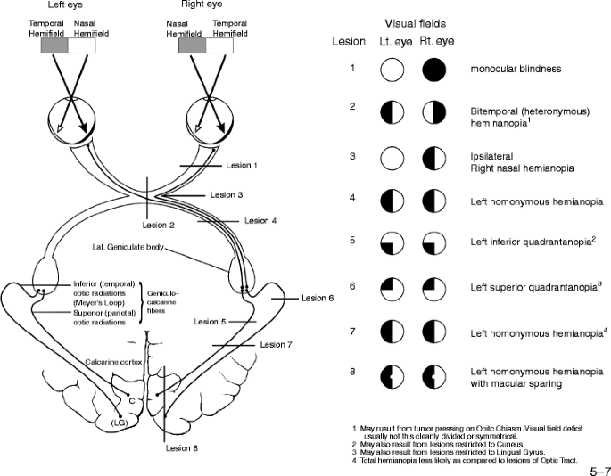
Clinical examination revealed only a BP - 150/90, R arm, sitting position with confrontation perimetry showing an incongruent, incomplete L sided homonymous hemianopia.
A homonymous hemianopia localizes posterior to the optic chiasma where the nasal hemiretinal fibers decussate.

A L sided HH localizes a lesion to the R post-chiasmatic visual pathway.

Incongruency argues for a relatively anterior localization of the lesion ie away from cortex. Image
Read 10 tweets
Jun 10, 2023
A lot of referrals to neurology are basically what I like to call 'lazy' referrals.

For example, you get a patient with paraparesis and instead of performing a detailed clinical evaluation, you shotgun some MRI and NCS and send a quick referral to neurology.
Since these investigations are poorly chosen and poorly aimed, the end result is mass confusion where localization goes for a toss.

General medicine has been particularly egregious in this regard.
If your knee jerk reaction is to just get an MRI LS spine for every low back pain, don't be surprised when the patient then develops an LMN type of lower limb weakness. Low back pain can be severe in GBS.
Read 6 tweets
Apr 16, 2023
I have always been tremendously interested in the history of medicine and its quirky stories.

One of the most interesting is West syndrome.

This is one of the rare syndromes which is simultaneously named after a physician and a patient.
In 1841, the general practitioner W.J. West from Turnbridge wrote a letter to the Lancet entitled 'on a peculiar form of infantile convulsions' describing symptomatology in his own son.
Image
Image
William James West was born im 1794 and became a member of the Royal College of Surgeons in 1815.

His 'son' was James Edwin, born in 1840 and transferred to the Earlwood Asylum for the Feeble-Minded in Redhill in 1853. He died 7 years later and was buried with his father.
Read 6 tweets
Mar 30, 2023
Interesting case in the ward today!

37y male first presented 10 yrs back, with insidious onset, progessively worsening R loss of vision --> complete blindness by 6 months.

He now presents with similar involvement of the L eye.

No other neurological/systemic hx.
On exam,

Gen survey - NAD

Neuro exam

CN 2 - R sided only perception of light, L finger count at 4 feet with colour desat, fundoscopy - B/L optic disc pale, L eye temporal hemianopia

CN 3,4,6 - EOM full in all directions, both pupils mid dilated and sluggishly R to light
Rest of exam is NAD.

What is your localization and pathology?

#MedTwitter
#neurotwitter
Read 11 tweets

Did Thread Reader help you today?

Support us! We are indie developers!


This site is made by just two indie developers on a laptop doing marketing, support and development! Read more about the story.

Become a Premium Member ($3/month or $30/year) and get exclusive features!

Become Premium

Don't want to be a Premium member but still want to support us?

Make a small donation by buying us coffee ($5) or help with server cost ($10)

Donate via Paypal

Or Donate anonymously using crypto!

Ethereum

0xfe58350B80634f60Fa6Dc149a72b4DFbc17D341E copy

Bitcoin

3ATGMxNzCUFzxpMCHL5sWSt4DVtS8UqXpi copy

Thank you for your support!

Follow Us!

:(