Mitral inflow Doppler and LVOT VTI in tamponade.
'ALT' for description #POCUS#echofirst#MedEd
Pulsus paradoxus: during inspiration, right heart filling occurs at the expense of the left, so that its transmural pressure transiently improves & then reverts during expiration (Ventricular interdependence). Seen as 👆on #POCUS
#1 Let's start with basics
Color Doppler identifies the flow + tells the direction (blue is away & red towards the probe [BART])
#2 👆BART holds good unless u invert the scale.
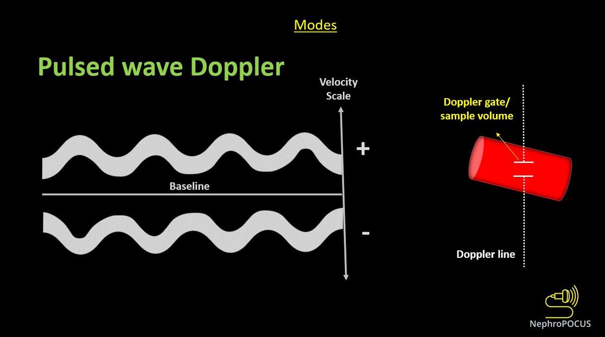
👇Pulsed wave Doppler (PWD) depicts blood flow at a certain point (sample volume) - we analyze the pattern of flow + velocity using this mode.
Above-the-baseline = flow towards the probe (like red on color)
Below = away (like blue)
#3
While performing any Doppler study, it's important to keep in mind that the angle between US beam & blood flow determines the accuracy of velocity displayed. Parallel = best, perpendicular = worst
As #VExUS does not rely on absolute velocities, its OK not to have perfect angle
#POCUS#echofirst quiz:
In this PLAX view, what structure does the arrow indicate? + what's abnormal about it (if any)?
Clinical: 71y pt with fever/cough, initially thought to be pneumonia, later developed chest pain. BP 134/83 Pulse 106
Will post the answer/source tonight #MedEd
Tweet 1/3
Answer:
Dilated coronary sinus with a mobile echogenicity (vegetation/infective endocarditis in this case)
Full case: 🔗ncbi.nlm.nih.gov/pmc/articles/P…
From #POCUS standpoint, 2 main causes: persistent left superior vena cava & pulmonary HTN. This pt had the former.
2/3 Normally on PLAX view, coronary sinus is barely visible. But when dilated, it can be confused with descending aorta (esp. by users who never encountered this scenario)
One of my favorite topics in #POCUS - various effusions seen from different sonographic windows.
Here is a thread 🧵with labeled images for those interested. 1. PLAX view - pericardial effusion - anterior to descending aorta ⚫️below LA), wedge shaped #MedEd#IMPOCUS#Nephpearls
2/ PSAX papillary muscle view
LV is think as many of my patients have #CKD and LVH (as well as mitral annular calcification seen on PLAX view above) #Nephrology#POCUS
3/ Apical 4-chamber view
Sometimes, effusions may be missed due to inadequate visualization of ventricular free wall/lung interference. Always pay attention to the RA area #POCUS
Looks like #POCUS ologists are in a mood to revive old #VExUS posts and tweetorials today.
Let me re-share the VExUS flash card(s) 🧵 1. VExUS grading live card #MedEd#IMPOCUS
@Rajiv_Sinanan@ThinkingCC 1/ Real reason(s) based on my conversations with people from different backgrounds:
Private practice: "#POCUS sounds interesting but I ain't got time for that. Will consider if I get compensated"
Academia: (not generalizing but most places)
Emphasis is on funded research
@Rajiv_Sinanan@ThinkingCC 2/ Clinical faculty are hired for clinical duties, outreach dialysis etc., which is fair but the problem is educational endeavors are rarely rewarded/provided with protected time. Faculty who want to be #POCUS champions must first learn it (either from institutional experts or
@Rajiv_Sinanan@ThinkingCC 3/ external courses), then integrate into curriculum, take care of administrative stuff/image archiving, prepare didactic material etc., which consume a lot of time and effort. Without dedicated protected time, its almost impossible to continue the initial enthusiasm.