ICU POCUS snippets: A bit of context: An elderly patient with hx of DM2 / HTN / HLD / peripheral vascular disease / ureteral stent & recurrent UTIs is admitted to the hospitalists’ service w diffuse abdominal pain, nausea & vomiting. Treated for a few days w antibiotics...
...but never really felt any better (weak/abd pain). Eventually, became hypotensive & was transferred to the ICU for “initiation of vasopressors”. Phys exam: diffuse abd tenderness. Formal echo earlier that day: "Normal LV/RV in size and systolic function". ICU POCUS was done...
...to gain more information regarding the cause of the abd pain and the hemodynamic picture. Some of the clips are shown here:
What is this 👆?
This is actually the pulmonary artery bifurcation. Sometimes u can even see a saddle pulmonary embolus sitting there... We usually get this view from the parasternal short axis at the AoV level aiming a bit higher. Here we were lucky to get it from a poor parasternal long axis:
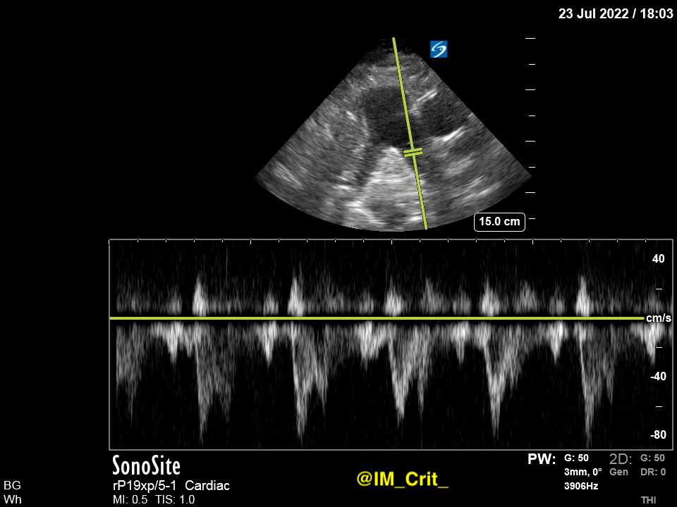
It is not always as clear as in this case but, if it is, you can interrogate w color Doppler and PW/CW separately each one of the PA branches (R - L):
PW Doppler in one side:
Sometimes you can see different PW profiles in the 2 branches... 👇 👇
PW Doppler focusing on the other side:
How is this 👆Doppler profile of the PA artery called? @khaycock2 has a great talk on this
Yes, I know, you are wondering how all these clips changed patient's management. They didn't, mainly because a "goals of care" discussion shifted the focus of our efforts. Next time they will. And, wouldn't it be nice to make our practice less boring?
ICU (evolving) stories: A young patient was admitted with "aspiration pneumonia" a few days ago. On mechanical ventilation. Afebrile. Negative cultures. CXR when you first see him (ET tube a bit deep, by the way):
You take a look at the ventilator screen. Patient on assist/volume control, 25 breaths, Vt 300 cc, FiO2 80%, PEEP 5.
U are a strong believer of guideline-directed medical therapies (GDMT). U know that following the PEEP table - as used in the ARDSnet study (NEJM 2004; 351(4): 327-36.
doi: 10.1056/NEJMoa032193) - is a well-tested way to set PEEP. For FiO2 of 80%, the recommended PEEP is:
ICU POCUS snippets: Much has been said about how useful lung POCUS is for procedural guidance. First of all, it accurately reveals large effusions when the radiology report characterizes them as “small”. This is from a recent case of a pt intubated w community-acquired pneumonia
and what proved to be bilateral parapneumonic effusions:
Secondly, while the dogma (which, btw, I don’t recommend completely ignoring!) in thoracentesis is to insert the needle at the “triangle of safety”, bordered by the anterior border of the latissimus dorsi, the lateral border of the pectoralis major, the horizontal line at the...
ICU quiz: A middle-aged patient w PMHx of COPD/neck Ca/lung Ca with a questionable L mainstem endobronchial lesion is in your ICU with resp failure. Doing "ok" on non-invasive ventilation for a couple of days but last night he was intubated. His CXR looks like this:
He is on VCV 360 cc x18 / peep 6 / fio2 60% w O2 sat 98% & Paco2 50 (pH 7.35). His ideal body weight: 60 kg. Pplat: 27, Ppeak: 23; there is no auto-PEEP. "Looks comfortable on the vent" breathing 18/min. Your bronchoscope is broken. What ventilator changes would u make (if any)?
ICU/CCU/Pharmacy pearls: Adenosine is another one of my favorite drugs (again: no COI); who doesn’t want to walk into a patient’s ward room after a rapid response is called for a HR of 190/min, administer 6 mg of adenosine and head back to the ICU 10 min later leaving the patient
on SR 80/min and the ICU charge nurse relieved that she will not have to find a creative way to “open up” another ICU room. Adenosine push is one of the VERY FEW intensivists’ triumphant moves, so I will take it. Nevertheless, there are a few things about adenosine use
that I think are fun or good to know (there are probably more than few, I just don't know them!): 1. Adenosine is a natural substance formed by the degradation of adenosine triphosphate (ATP); yes, that ATP! So, in theory
ICU stories: You start your night shift and while walking in and out each patient’s room, you see this 👇 on one ventilator's screen:
The patient (I know: I should have looked at the patient first, not at the ventilator screen... 🤷♂️) is breathing like this 😳:
Quick chart review: middle-aged pt admitted w ARDS > 1 month ago. Already w tracheostomy + PEG. Still unable to be weaned on trach mask, despite being on "moderate" fio2 of 40-50%. On iv sedation; drowsy, hemodynamically stable. Not febrile or acidotic. No "weird" labs. CXR:
Several of my colleagues living/working outside the United States are surprised to learn that:
1. Many US hospitals have intensive care units but no intensivists. This is unimaginable in many European countries
2. Many US hospitals (even medium-sized with 200 beds) have no surgeon or cardiologist or anesthesiologist (or their respective specialty trainees) in-house at night-time or during the weekend
3. Many US hospitals have only 3 physicians in-house during the night shift: an emergency medicine, an internal medicine / family medicine (hospitalist) and an intensivist with/without help from physician assistants