#POCUS quiz for #VExUS enthusiasts.
Image obtained from a patient with heart failure with preserved EF. IVC 1.9 cm with 30% inspiratory collapse.
Here is the intra-renal image. Interpretation of the venous waveform?
POLL in thread 👇 #MedEd#Nephrology
👆#POCUS
S = systolic venous wave
D = diastolic venous wave
CVP waveform - a variant of #VExUS#POCUS
Tracings obtained from a 38-year-old female with severe aortic stenosis and mild pulmonary hypertension who underwent aortic valve replacement. What's the difference from pre to post-op?
Se thread for answer and source. #MedEd
Mitral inflow Doppler and LVOT VTI in tamponade.
'ALT' for description #POCUS#echofirst#MedEd
Pulsus paradoxus: during inspiration, right heart filling occurs at the expense of the left, so that its transmural pressure transiently improves & then reverts during expiration (Ventricular interdependence). Seen as 👆on #POCUS
#1 Let's start with basics
Color Doppler identifies the flow + tells the direction (blue is away & red towards the probe [BART])
#2 👆BART holds good unless u invert the scale.
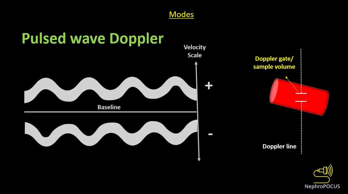
👇Pulsed wave Doppler (PWD) depicts blood flow at a certain point (sample volume) - we analyze the pattern of flow + velocity using this mode.
Above-the-baseline = flow towards the probe (like red on color)
Below = away (like blue)
#3
While performing any Doppler study, it's important to keep in mind that the angle between US beam & blood flow determines the accuracy of velocity displayed. Parallel = best, perpendicular = worst
As #VExUS does not rely on absolute velocities, its OK not to have perfect angle
#POCUS#echofirst quiz:
In this PLAX view, what structure does the arrow indicate? + what's abnormal about it (if any)?
Clinical: 71y pt with fever/cough, initially thought to be pneumonia, later developed chest pain. BP 134/83 Pulse 106
Will post the answer/source tonight #MedEd
Tweet 1/3
Answer:
Dilated coronary sinus with a mobile echogenicity (vegetation/infective endocarditis in this case)
Full case: 🔗ncbi.nlm.nih.gov/pmc/articles/P…
From #POCUS standpoint, 2 main causes: persistent left superior vena cava & pulmonary HTN. This pt had the former.
2/3 Normally on PLAX view, coronary sinus is barely visible. But when dilated, it can be confused with descending aorta (esp. by users who never encountered this scenario)