Lea Alhilali, MD Profile picture
Oct 7, 2022 16 tweets 9 min read Read on X
1/When it comes to brachial plexus pathology, you shouldn’t have to wing it! A #tweetorial to help you figure out #brachialplexus injuries
#medtwitter #meded #neurosurgery #orthotwitter #orthopedics #neurorad #radres #medstudent #FOAMed #FOAMrad #spine #radiology #neurotwitter Image
2/When it comes to brachial plexus injuries, the most important distinction to make is whether it is a pre or post ganglionic injury. Pre ganglionic injuries are nerve root avulsions from the spinal cord and cannot be repaired, while most post ganglionic injuries can be repaired Image
3/It’s like repairing a house. If there’s a structural flaw at the beginning, before you’ve even built it, you won’t even try to build it, you’ll just scrap the design (preganglionic). But if there’s damage after the house is built, you can salvage it w/repair (postganglionic) Image
4/Preganglionic injuries often have an associated pseudomeningocele. This is bc when you rip the nerve root out, you also rip the dura—like pulling the cork out of a champagne bottle—and CSF will leak, creating a collection. Image
5/Another sign of a preganglionic injury is that there is no identifiable nerve root. B/c the nerve root has been ripped out and retracted, you will not see it in the thecal sac. The nerve root sleeve will also be empty—an empty shell of where the nerve root used to be. Image
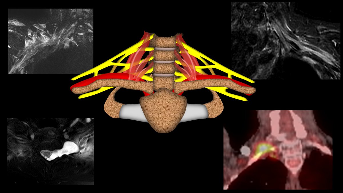
6/For plexus injuries, remember, the plexus is how cervical nerve roots jump down to the arm—kind of like a bungee jumper—tied to the top while jumping down to the bottom. So all the bad things that can happen to a bungee jumper can help us remember what harms the brachial plexus Image
7/First problem on the way down—the bungee cord attachment rips off—and you are left free falling 😬. This is like a preganglionic injury. And just like what happens to the jumper when the cord attachment rips off--there is no salvage w/this type of injury Image
8/Remember, for preganglionic injuries, look to see if the nerve root is missing and look for the characteristic pseudomeningocele. Image
9/Next problem on the way down—getting hit by external objects flying through the air. This is like radiation therapy—it comes flying from outside to hit the brachial plexus. Image
10/Radiation plexopathy will show long segment or diffuse thickening or enhancement of the plexus, but there won’t be an identifiable focal mass. Where you see the thickening depends on the cancer treated. Head & neck XRT will affect higher, while lung XRT will affect lower. Image
11/Next problem on the way down—crashing into something in the vicinity. This is like trauma. The brachial plexus can crash into the clavicle in front of it, like crashing into the rocks below. It can also be stretched as well. Image
12/Three main things to look for w/traumatic plexus injury. (1) Edema will usually indicate injury. (2) Thickening could hide a neuroma in continuity, so it needs follow up, (3) Discontinuity—nerve is disrupted. Postganglionic disruptions can possibly be repaired w/microsurgery Image
13/Where the injury occurs determines if it can be repaired. The Nagano injury zones show that while we knew we couldn’t repair preganglionic injuries, post ganglionic injuries not past the foramen are too difficult to repair. So if it’s not out of the foramen, it’s foregone! Image
14/Finally, the jumper can get eaten by something—this is involvement by tumor. It can be an intrinsic tumor like a schwannoma, or metastases from something like breast, but most commonly, it is direct invasion by a Pancoast lung tumor. Image
15/A tumor can be differentiated from radiation changes by seeing a focal mass. Because Pancoast lung tumors come from below, they usually affect the lower nerve roots, C7 and T1. Image
16/So now you can remember the brachial plexus pathologies by remembering the unfortunate things that can happen to a bungee jumper on the way down. So while brachial plexus injuries may cause winging—when it comes to diagnosing these injuries, you won’t have to wing it! Image

• • •

Missing some Tweet in this thread? You can try to force a refresh
 

Keep Current with Lea Alhilali, MD

Lea Alhilali, MD Profile picture

Stay in touch and get notified when new unrolls are available from this author!

Read all threads

This Thread may be Removed Anytime!

PDF

Twitter may remove this content at anytime! Save it as PDF for later use!

Try unrolling a thread yourself!

how to unroll video
  1. Follow @ThreadReaderApp to mention us!

  2. From a Twitter thread mention us with a keyword "unroll"
@threadreaderapp unroll

Practice here first or read more on our help page!

More from @teachplaygrub

Jun 27
1/Blast from the past!

Sometimes to be next gen, you gotta to go old school!

Cutting edge pituitary imaging must be MRI, right?

Or can we go back to the future w/CT?

Here’s the latest in pituitary imaging in this month’s @theAJNR SCANtastic!

ajnr.org/content/45/6/7…
Image
2/Pituitary imaging is actually very difficult.

First challenge is the small size of the gland & even smaller adenomas, requiring high resolution.

And the difference between adenomas & the gland is subtle—both enhance, but adenomas enhance SLIGHTLY less Image
3/The difference in enhancement is transient & ends quickly

So pituitary imaging must be done dynamically to catch this small window of difference

So we have to do very high resolution imaging very quickly—the worst of both worlds! Image
Read 12 tweets
Jun 21
1/”I LOVE spinal cord syndromes!” is a phrase that has NEVER, EVER been said by anyone.

Do you become paralyzed when you see cord signal abnormality?

Never fear—here is a thread on all the incomplete spinal cord syndromes to get you moving again! Image
2/Spinal cord anatomy can be complex.

On imaging, we can see the ant & post nerve roots.

We can also see the gray & white matter.

Hidden w/in the white matter, however, are numerous efferent & afferent tracts—enough to make your head spin. Image
3/Lucky for you, for the incomplete cord syndromes, all you need to know is gray matter & 3 main tracts

Anterolaterally, spinothalamic tract (pain & temp). Posteriorly, dorsal columns (vibration, proprioception, & light touch), & next to it, corticospinal tracts—providing motor Image
Read 20 tweets
Jun 19
1/”Tell me where it hurts.”

How back pain radiates can tell you where the lesion is—if you know where to look!

Remembering lumbar radicular pain distributions can be back breaking work--but here's a thread to help you! Image
2/Let’s start with L1.

L1 radiates to the groin.

I remember that b/c the number 1 is, well, um…phallic.

So the phallic number 1 radiates to the groin. Image
3/Let’s skip to L3 for a second.

I remember L3 is to the knee—easy, it rhymes! Image
Read 8 tweets
Jun 10
1/Do you know all the aspects of, well, ASPECTS?

Many know the anterior circulation stroke system—but posterior circulation (pc) ASPECTS is often left behind

25% of infarcts are posterior circulation

Do you know pc-ASPECTS?!

Here’s a thread to help you remember pc-ASPECTS Image
2/Many know anterior circulation ASPECTS.

It uses a 10-point scoring system to semi-quantitation the amount of the MCA territory infarcted on non-contrast head CT

If you need a review: here’s my thread on ASPECTS:
Image
3/But it’s only useful for the anterior circulation.

Posterior circulation accounts for ~25% of infarcts

Even w/recanalization, many of these pts do poorly bc of the extent of already infarcted tissue

So there’s a need to quantitate the amount of infarcted tissue in these pts Image
Read 8 tweets
May 29
1/Waving the white flag when it comes to white matter anatomy?

Turns out white matter anatomy isn’t black & white!

This months @theAJNR SCANtastic is the white knight you need to rescue you!

Here’s the white matter anatomy you NEED to know!

ajnr.org/content/45/5/5…
Image
2/Gray matter or cortical functional anatomy is well known.

Everyone knows the motor & sensory strips. Most know Broca’s & Wernicke’s

But most forget that white matter has similar functional topography & just bc it’s white matter doesn’t mean it doesn’t have function! Image
3/But too often we don’t refer to this white matter functional anatomy.

Instead we use general terms like “corona radiata”

But that’s the equivalent of using the word “body.”

Just like the body has many different systems in it, so does the corona radiata! Image
Read 12 tweets
May 21
1/Having trouble remembering what you should look for in vascular dementia on imaging?

Almost everyone worked up for dementia has infarcts. Which ones are important?

Here’s a thread on the key findings in vascular dementia! Image
2/Vascular cognitive impairment, or its most serious form, vascular dementia, used to be called multi-infarct dementia.

It was thought dementia directly resulted from brain volume loss from infarcts, w/the thought that 50-100cc of infarcted related volume loss caused dementia Image
3/But that’s now outdated. We now know vascular dementia results from diverse pathologies that all share a common vascular origin.

It’s possible to lose little volume from infarct & still result in dementia.

So if infarcts are common—which contribute to vascular dementia? Image
Read 21 tweets

Did Thread Reader help you today?

Support us! We are indie developers!


This site is made by just two indie developers on a laptop doing marketing, support and development! Read more about the story.

Become a Premium Member ($3/month or $30/year) and get exclusive features!

Become Premium

Don't want to be a Premium member but still want to support us?

Make a small donation by buying us coffee ($5) or help with server cost ($10)

Donate via Paypal

Or Donate anonymously using crypto!

Ethereum

0xfe58350B80634f60Fa6Dc149a72b4DFbc17D341E copy

Bitcoin

3ATGMxNzCUFzxpMCHL5sWSt4DVtS8UqXpi copy

Thank you for your support!

Follow Us!

:(