Lea Alhilali, MD Profile picture
Nov 4, 2022 15 tweets 8 min read Read on X
1/Hate it when one radiologist called the stenosis mild, the next one said moderate--but it was unchanged?!

Here’s a #tweetorial of a lumbar grading system that’s easy, reproducible & evidence-based

#medtwitter #spine #neurosurgery #radres #neurorad #meded #FOAMed #FOAMrad Image
2/Lumbar stenosis has always been controversial. In 2012, they tried to survey spine experts to come to a consensus as to what are the most important criteria for canal & foraminal stenosis. And the consensus was…that there was no consensus. So what should you use to call it? Image
3/Well, you don’t want just gestalt it—that is a recipe for inconsistency & disagreement. But you don’t want to measure everything either—measurements are not only cumbersome, they introduce reader variability & absolute measurements don’t mean the same thing in every patient. Image
4/Think of it functionally. Nerves need to fit in their space, like you fit in clothing. Mild stenosis is like comfy clothes—no squeezing. Moderate stenosis clothing isn’t loose, but there isn’t extra room either. Severe stenosis is like too tight jeans, your body gets compressed Image
5/So how do we tell if the nerves have enough room—if the clothing fits loosely, tight, or too tightly? We look at the space around them. For the canal, it is CSF—if there's enough room, extra space will be filled by CSF. For foramina, it is fat—extra room is filled by fat. Image
6/For mild canal stenosis, there is mild attenuation of the CSF space, but there is still plenty of CSF around, just like there is plenty of room in your comfy sweat pants Image
7/For moderate canal stenosis, the canal starts closing in, so there is less CSF around and the nerve roots appear aggregated. It’s like the clothes you wear to the club, there isn’t much room between your skin & the clothing, but you can still fit into them (hopefully) Image
8/Severe canal stenosis is the too tight jeans. The canal doesn’t just hug up to the nerve roots, it compresses them. Like your belly after a big meal trying to get into tight jeans, they get squished and deformed in order to fit, so they can’t be separated from each other. Image
9/This classification isn’t just easy to remember, it’s also evidence based. This is the Lee classification that has excellent reproducibility not just among radiologists, but among everyone. And it does correlate w/increasing symptoms. Image
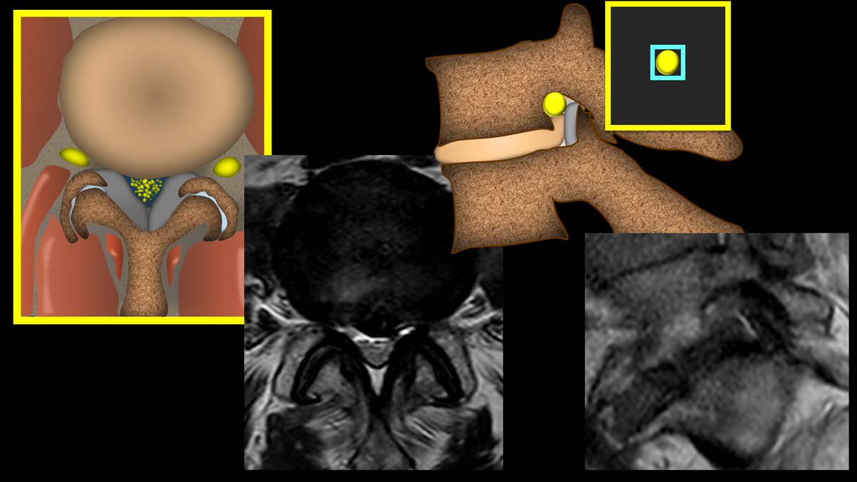
10/For foraminal narrowing, the nerve inside the foramen has fat around it on four sides that can be attenuated as the space gets tighter. How many sides are attenuated determines how severe the stenosis is. Image
11/Mild stenosis is where you have loss of the fat on 2 sides. So it is still comfy clothing bc the fat is preserved on the other two sides, so you still have lots of space. Image
12/For moderate stenosis, you lose the space on all four sides, but the nerve itself is not compressed or deformed. Like a sleek outfit, it shows your curves, but doesn’t deform them. It’s not a comfy outfit, per se, & I wouldn’t eat a lot while wearing it, but it’s not too small Image
13/For severe stenosis, we are trying to fit into those jeans from high school and it isn’t going well. You are squishing in everything you can to get it to fit. Same with the foramen—the fat isn’t just gone, the nerve is compressed and deformed. Image
14/This is also named the Lee system. It fits well with the Lee classification for canal stenosis. It also is extremely reproducible and correlates with findings at surgery.

So you don’t have to remember a complicated system—just ask yourself, how does the clothing fit? Image
15/So put away your measuring calipers! You can end the inter-observer variability. These systems are easy to remember, make sense, and are based in evidence. As they say, if it fits—wear it! Image

• • •

Missing some Tweet in this thread? You can try to force a refresh
 

Keep Current with Lea Alhilali, MD

Lea Alhilali, MD Profile picture

Stay in touch and get notified when new unrolls are available from this author!

Read all threads

This Thread may be Removed Anytime!

PDF

Twitter may remove this content at anytime! Save it as PDF for later use!

Try unrolling a thread yourself!

how to unroll video
  1. Follow @ThreadReaderApp to mention us!

  2. From a Twitter thread mention us with a keyword "unroll"
@threadreaderapp unroll

Practice here first or read more on our help page!

More from @teachplaygrub

Mar 16
1/Remembering spinal fracture classifications is back breaking work!

A thread to review the scoring system for thoracic & lumbar fractures—“TLICS” to the cool kids! Image
2/TLICS scores a fx on (1) morphology & (2) posterior ligamentous complex injury

Let's start w/morphology

TLICS scores severity like the steps to make & eat a pizza:

Mild compression (kneading), strong compression (rolling), rotation (tossing), & distraction (tearing in) Image
3/At the most mild, w/only mild axial loading, you get the simplest fx, a compression fx—like a simple long bone fx--worth 1 pt.

This is like when you just start to kneading the dough. There's pressure, but not as much as with a rolling pin! Image
Read 13 tweets
Mar 14
1/The 90s called & wants its carotid imaging back!

It’s been 30 years--why are you still just quoting NASCET?

Do you feel vulnerable when it comes to identifying plaque vulnerability?

Here’s a thread to help you identify high risk plaques with carotid plaque imaging Image
2/Everyone knows the NASCET criteria:

If the patient is symptomatic & the greatest stenosis from the plaque is >70% of the diameter of normal distal lumen, patient will likely benefit from carotid endarterectomy.

But that doesn’t mean the remaining patients are just fine! Image
3/Yes, carotid plaques resulting in high grade stenosis are high risk.

But assuming that stenosis is the only mechanism by which a carotid plaque is high risk is like assuming that the only way to kill someone is by strangulation. Image
Read 25 tweets
Mar 12
1/Do you know all the aspects of, well, ASPECTS?

Many know the anterior circulation stroke scoring system—but posterior circulation (pc) ASPECTS is often left behind

25% of infarcts are posterior circulation

Do you know pc-ASPECTS?!

Here’s how to remember pc-ASPECTS! Image
2/Many know anterior circulation ASPECTS.

It uses a 10-point scoring system to semi-quantitation the amount of the MCA territory infarcted on non-contrast head CT

If you need a review: here’s my thread on ASPECTS: Image
3/But it’s only useful for the anterior circulation.

Posterior circulation accounts for ~25% of infarcts.

Even w/recanalization, many of these pts do poorly bc of the extent of already infarcted tissue.

So there’s a need to quantitate the amount of infarcted tissue in these ptsImage
Read 12 tweets
Mar 10
1/I always say you can tell a bad read on a spine MR if it doesn’t talk about lateral recesses.

What will I think when I see your read? Do you rate lateral recess stenosis?

Here’s a thread on lateral recess anatomy & a grading system for lateral recess stenosis Image
2/First anatomy.

Thecal sac is like a highway, carrying the nerve roots down the lumbar spine.

Lateral recess is part of the lateral lumbar canal, which is essentially the exit for spinal nerve roots to get off the thecal sac highway & head out into the rest of the body Image
3/Exits have 3 main parts.

First is the deceleration lane, where the car slows down as it starts the process of exiting.

Then there is the off ramp itself, and this leads into the service road which takes the car to the roads that it needs to get to its destination Image
Read 21 tweets
Mar 3
1/Does PTERYGOPALATINE FOSSA anatomy feel as confusing as its spelling?

Does it seem to have as many openings as letters in its name?

Are you pterrified of the pterygopalatine fossa (PPF)?

Let this thread on PPF anatomy help you out. Image
2/The PPF is a crossroads between the skullbase & the extracranial head and neck

There are 4 main regions that meet here:

(1) Skullbase itself posteriorly, (2) nasal cavity medially, (3) infratemporal fossa laterally, and (4) orbit anteriorly. Image
3/At its most basic, you can think of the PPF as a room with 4 doors opening to each of these regions: one posteriorly to the skullbase, one medially to the nasal cavity, one laterally to the infratemporal fossa, and one anteriorly to the orbit Image
Read 18 tweets
Feb 28
1/Feel like a fish out of water when it comes to water on the brain?

Read on for this month’s @Radiographics summary of what you need to know about hydrocephalus!!


@cookyscan1 @RadG_editor #RGphx doi.org/10.1148/rg.240…Image
2/To understand hydrocephalus, think of CSF like the flow of traffic

3 main ways traffic backs up:

(1) Obstruction on the road:
For hydrocephalus, this is an obstruction along CSF in the ventricle Image
3/

(2) Obstruction of an off ramp
For hydrocephalus=obstruction at its off ramp into the venous system

(3) Rush hour
For hydrocephalus=over production Image
Read 8 tweets

Did Thread Reader help you today?

Support us! We are indie developers!


This site is made by just two indie developers on a laptop doing marketing, support and development! Read more about the story.

Become a Premium Member ($3/month or $30/year) and get exclusive features!

Become Premium

Don't want to be a Premium member but still want to support us?

Make a small donation by buying us coffee ($5) or help with server cost ($10)

Donate via Paypal

Or Donate anonymously using crypto!

Ethereum

0xfe58350B80634f60Fa6Dc149a72b4DFbc17D341E copy

Bitcoin

3ATGMxNzCUFzxpMCHL5sWSt4DVtS8UqXpi copy

Thank you for your support!

Follow Us!

:(