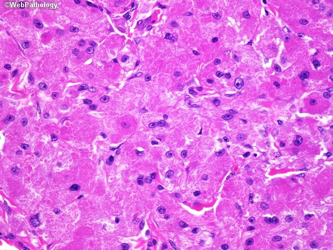
A 48 year old male presented with submandibular swelling for 3 weeks.On examination the swelling was painless, solid, firm and immobile. Excision was performed. Hand E stained section of the tumour is shown below. What is your diagnosis? #pathtweetorial #pathboards #pathresidents 

Options:
Answer is Acinic cell carcinoma -"acinic cells" (abundant finely vacuolated cytoplasm with basophilic granules, small nuclei with stippled chromatin), scattered "intercalcated duct type cells" (eosinophilic cytoplasm with moderate amount of cytoplasm and bland nuclei).

Basophilic granules found in acinic cell carcinoma are PAS positive and Diastase resistant. Same pattern of staining can be observed in the granules of which other tumor?
Answer is Granular cell tumor
The granules are eosinophilic in granular cell tumor, PAS-positive and diastase resistant.
Ewings sarcoma has PAS-poitive material which is sensitive to diastase.
The granules are eosinophilic in granular cell tumor, PAS-positive and diastase resistant.
Ewings sarcoma has PAS-poitive material which is sensitive to diastase.
Last question on tumors with cytoplasmic granules and crystals.
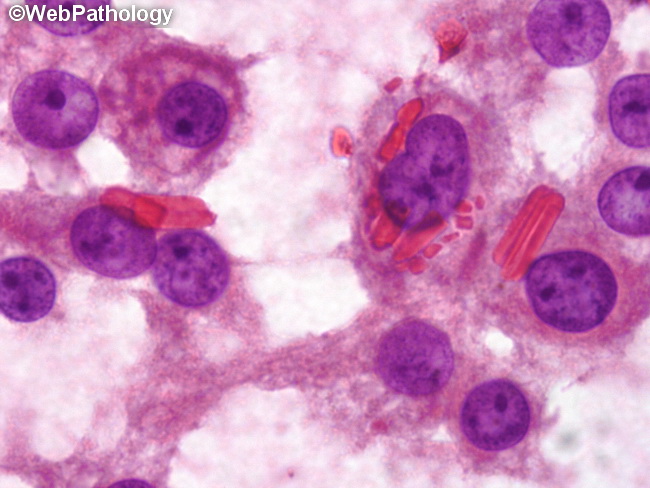
Which tumor is likely to show cytoplasmic eosinophilic crystals (image)
Which tumor is likely to show cytoplasmic eosinophilic crystals (image)
Options:
Answer is Leydig cell tumor . Reinke's crystalloids (cytoplasmic eosinophilic crystals) are seen in about 30-40% of cases and appear as dense needle-like or rhomboid structures in the cytoplasm.
Mention some tumors with cytoplasmic granules.
Thanks .
Mention some tumors with cytoplasmic granules.
Thanks .
• • •
Missing some Tweet in this thread? You can try to
force a refresh