
1/ Let’s talk about P waves #CardioTwitter.
The P wave is the first positive deflection on the EKG and represents atrial depolarization. The first half represents right atrial depolarization and the second half represents left atrial depolarization.
By @ekgdx
The P wave is the first positive deflection on the EKG and represents atrial depolarization. The first half represents right atrial depolarization and the second half represents left atrial depolarization.
By @ekgdx

2/ Normal P wave
Criteria
✅ Axis: 0° to +75°.
✅ Amplitude (L leads): <2.5 mm.
✅ Amplitude (P leads): <1.5 mm.
✅ Duration: 0.08 - 0.11 sec.
✅ Morphology: Upright in I, II, aVF and inverted in aVR.
#Pwaves #ecg #ekg #ekgdx #medicine #MedTwitter #MedStudentTwitter #basic
Criteria
✅ Axis: 0° to +75°.
✅ Amplitude (L leads): <2.5 mm.
✅ Amplitude (P leads): <1.5 mm.
✅ Duration: 0.08 - 0.11 sec.
✅ Morphology: Upright in I, II, aVF and inverted in aVR.
#Pwaves #ecg #ekg #ekgdx #medicine #MedTwitter #MedStudentTwitter #basic
3/ Peaked P wave
The morphology is peaked with amplitude ≥2.5 mm, usually in II, III and aVF.
The peaked P wave is a typical characteristic of right atrial abnormality/enlargement.
Causes: PE, COPD, congenital heart disease, pulmonary hypertension, normal variant, others.
The morphology is peaked with amplitude ≥2.5 mm, usually in II, III and aVF.
The peaked P wave is a typical characteristic of right atrial abnormality/enlargement.
Causes: PE, COPD, congenital heart disease, pulmonary hypertension, normal variant, others.

4/ Notched P wave
The morphology is notched like an "M" shape.
If the duration is ≥0.12 sec consider left atrial abnormality/enlargement.
Causes include: Heart failure, LVH, MI, mitral or aortic valve disease, others.
#ekgdx #ecg #medicine #MedTwitter #notched #pwave
The morphology is notched like an "M" shape.
If the duration is ≥0.12 sec consider left atrial abnormality/enlargement.
Causes include: Heart failure, LVH, MI, mitral or aortic valve disease, others.
#ekgdx #ecg #medicine #MedTwitter #notched #pwave

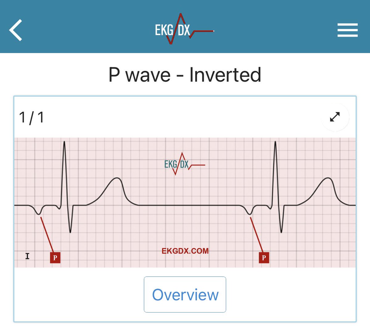
5/ Inverted P waves
Possible causes include:
✅ Ectopic atrial beat or rhythm.
✅ AV junctional premature complex or rhythm.
✅ Lead placement error.
✅ Dextrocardia.
✅ Others.
#ekgdx #medicine #MedTwitter #basic #ecg #ekg
Possible causes include:
✅ Ectopic atrial beat or rhythm.
✅ AV junctional premature complex or rhythm.
✅ Lead placement error.
✅ Dextrocardia.
✅ Others.
#ekgdx #medicine #MedTwitter #basic #ecg #ekg
6/ Retrograde P wave
The impulse can travel backward, in a retrograde fashion, through the atria, producing a retrograde P wave, as long as the impulse penetrate the AV node and depolarize the atria.
#retrograde #pwave
The impulse can travel backward, in a retrograde fashion, through the atria, producing a retrograde P wave, as long as the impulse penetrate the AV node and depolarize the atria.
#retrograde #pwave

7/ Retrograde P waves can be:
✅ Before the QRS.
✅ During the QRS.
✅ After the QRS.
Usually inverted in II, III and aVF and upright in aVR and V1.
✅ Before the QRS.
✅ During the QRS.
✅ After the QRS.
Usually inverted in II, III and aVF and upright in aVR and V1.
8/ Learn EKG using graphic explanation with a smart software that help you with the learning process.
The EKGs are courtesy of EKGDX.
You can download the App here: onelink.to/7bp23c
#CardioTwitter
The EKGs are courtesy of EKGDX.
You can download the App here: onelink.to/7bp23c
#CardioTwitter
11/ Let’s continue with the Q wave and QRS complex.
🧵👇
🧵👇
https://twitter.com/ekgdx/status/1595466662500831233
• • •
Missing some Tweet in this thread? You can try to
force a refresh


