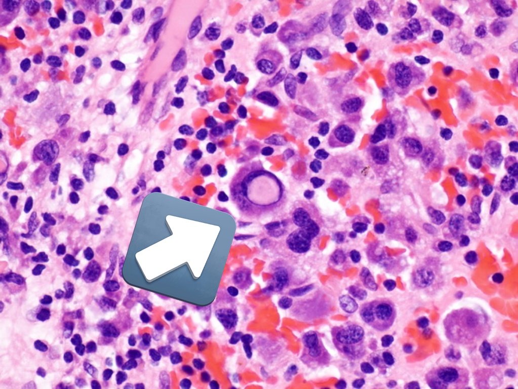
A 65 year old male presented with low urine output and bone pain. On further investigation serum creatinine was high and the patient had low hemoglobin. Bone marrow biopsy showed finding in the image. What is your diagnosis? #pathtwitter #pathtweetorial #pathresidents #pathboards 

Options
Answer is multiple myeloma.
The image shows "INTRA-NUCLEAR INCLUSIONS" called "DUTCHER BODIES".
They are deposits of immunoglobulins. Intracytoplasmic immunoglobulin inclusions are called RUSSEL BODIES.
Seen in multiple myeloma, plasmacytoma, lymphoplasmacytic lymphoma (LPL).
The image shows "INTRA-NUCLEAR INCLUSIONS" called "DUTCHER BODIES".
They are deposits of immunoglobulins. Intracytoplasmic immunoglobulin inclusions are called RUSSEL BODIES.
Seen in multiple myeloma, plasmacytoma, lymphoplasmacytic lymphoma (LPL).

Dutcher bodies are
Dutcher bodies are true inclusions.
True inclusions are due to deposition of substances such as immunoglobulins or viral particles, whereas pseudoinclusions are cytoplasmic invaginations
Image credit: mobile.twitter.com/megothelioma
True inclusions are due to deposition of substances such as immunoglobulins or viral particles, whereas pseudoinclusions are cytoplasmic invaginations
Image credit: mobile.twitter.com/megothelioma

Identify the odd man out ( All conditions mentioned below are true nuclear inclusions-except?)
Answer is Meningioma.
Intranuclear inclusions Seen in meningioma are "PSEUDO-INCLUSIONS"
Some conditions showing nuclear true inclusions are summarized in the images below.


Intranuclear inclusions Seen in meningioma are "PSEUDO-INCLUSIONS"
Some conditions showing nuclear true inclusions are summarized in the images below.


Last one on this topic.
Which tumor has characteristic intranuclear pseudo- inclusions?
Which tumor has characteristic intranuclear pseudo- inclusions?
Answer is all
Image shows some common tumors showing intranuclear inclusions.
Have you heard of pseudo-pseudo inclusions???
If you haven't, find out about it and leave a comment.
Thanks for staying!!
Image shows some common tumors showing intranuclear inclusions.
Have you heard of pseudo-pseudo inclusions???
If you haven't, find out about it and leave a comment.
Thanks for staying!!

• • •
Missing some Tweet in this thread? You can try to
force a refresh


