Daniel Gewolb, MD Profile picture
Dec 16, 2022 3 tweets 2 min read Read on X
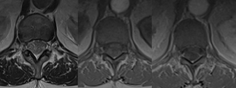
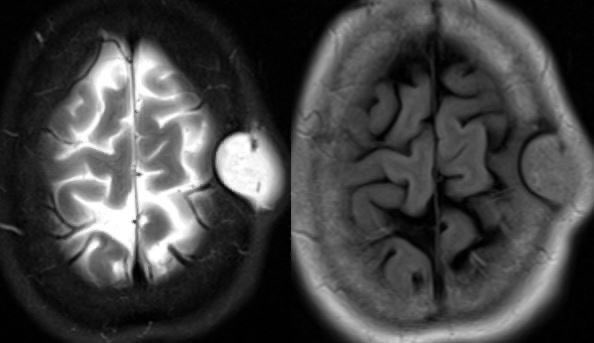
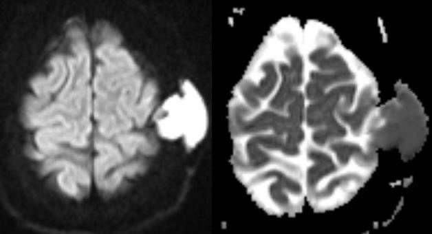
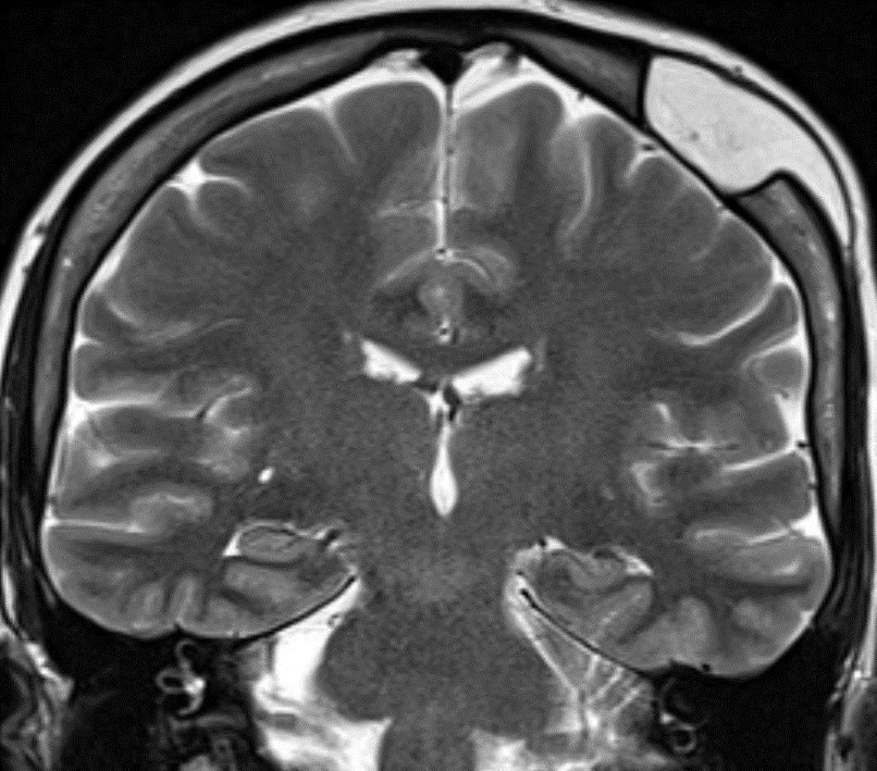
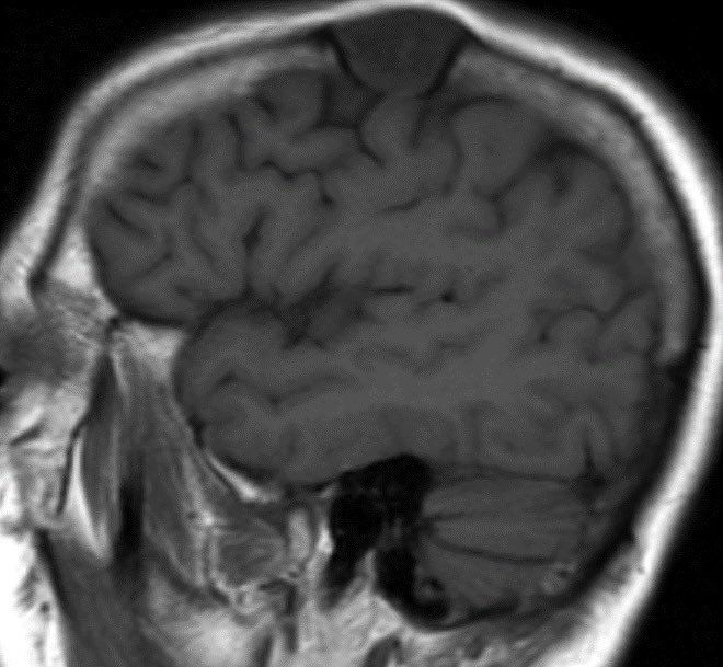
78 yr old with encephalopathy and seizure. MRI shows vasogenic edema within the left temporal and occipital white matter with subcortical extension. SWI shows characteristic lobar micro-hemorrhages predominantly in the effected regions.
#Neurology #neuroradiology #radres ImageImageImageImage
Ddx:
PML
Inflammatory amyloid angiopathy
PRES
Amyloid-related imaging abnormalities (ARIA)
Answer: Inflammatory amyloid angiopathy

This can be indistinguishable from amyloid-related imaging abnormalities (ARIA) seen in patients undergoing treatment for Alzheimer’s disease. Therapies include:

bapineuzumab, solanezumab and aducanumab

• • •

Missing some Tweet in this thread? You can try to force a refresh
 

Keep Current with Daniel Gewolb, MD

Daniel Gewolb, MD Profile picture

Stay in touch and get notified when new unrolls are available from this author!

Read all threads

This Thread may be Removed Anytime!

PDF

Twitter may remove this content at anytime! Save it as PDF for later use!

Try unrolling a thread yourself!

how to unroll video
  1. Follow @ThreadReaderApp to mention us!

  2. From a Twitter thread mention us with a keyword "unroll"
@threadreaderapp unroll

Practice here first or read more on our help page!

More from @daniel_gewolb

Aug 1
⭐️What is the most likely diagnosis in this patient presenting with a lump on the head?

More images in 🧵

#MedEd #medicine #neurology #neurosurgery #ENT #radres #peds @Radiopaedia #futureradres @AlbanyMedRadRes Image
Image
Image
Image
More images 👇 Image
Image
⭐️ Answer: Intradiploic epidermoid cyst

▶️CT shows a circumscribed low density skull mass with smooth scalloped margin suggesting a benign lesion

▶️Density/signal intensity is near CSF with the exception of FLAIR which shows incomplete suppression (dirty signal not black like CSF)Image
Image
Image
Image
Read 6 tweets
Jul 19
🔷What is the most likely diagnosis and clinical syndrome in this patient with left ear pain, cranial nerve 6 palsy, and retro-orbital pain? 🧠 🤔

#Neurology #ent #neurosurgery #radres #NeuroTwitter #futureradres #MRI #Medicine @ASHNRSociety @RSNA #Ophthalmology Image
Image
Image
Image
⭐️ Answer: petrous apicitis complicated by brainstem abscess

🔷Petrous apicitis can display the clinical triad of Gradenigo’s syndrome

1️⃣Otorrhea
2️⃣Cranial nerve 6 palsy
3️⃣Pain in distribution of trigeminal nerve
▶️Petrous apicitis usually occurs as a complication of otomastoiditis when the infection spreads to the skull base.

▶️The petrous apex is in close proximity to Dorello’s canal (where cranial nerve 6 runs through) and Meckel’s cave (where the trigeminal ganglion is located)
Read 6 tweets
Jul 4
⭐️ What is the most likely diagnosis in this pregnant patient presenting with seizures? 🧠

#Neurology #medicine #radres #futureradres #Neurosurgery #MRI #FOAMed Image
Image
Image
Image
⭐️ Answer: Cortical vein thrombosis (CVT)

▶️CVT causes retrograde venous pressure leading to focal vasogenic edema

▶️Increased back pressure is characterized by dilated veins and petechial hemorrhage which can progress to large hematomas and ischemic neurological damage
🔷Two types of edema can develop:
1️⃣Vasogenic (from venous back pressure)
2️⃣Cytotoxic (ischemia)

🔷Risk factors:
💡 Up to 20% are idiopathic
1️⃣Trauma
2️⃣Tumor/malignancy (compression/invasion from meningioma)
3️⃣Infection
4️⃣Hormonal (pregnancy)
5️⃣Dehydration
Read 7 tweets
Jun 22
🔷What is the most likely diagnosis in this 70 y/o F who lives with feral cats presenting w/ vomiting, diarrhea, leukocytosis, fever for 3 days and progressive decline in level of consciousness?

🔷CSF: initially normal, repeat a few days later ⬆️ WBC (lymphocyte predominant), ⬆️ Protein, normal glucoseImage
Image
Image
Image
⭐️ Answer: Viral encephalitis (Specifically Rabies)

🔷 South Central Asia and Southeast Asia appear to have the greatest number of cases

🔷Dogs are the most common reservoir 🐶 (bats in developed countries🦇)
🔷CLINICAL:

▶️Incubation period typically 3 weeks to 2 months (range 5 days to 6 months)
▶️Prodromal symptoms: fever, malaise, anxiety,
and itching at the inoculation site

💡Once in the body, the virus begins retrograde flow to extend to the dorsal root ganglion, which may correlate with neuropathic pain

▶️CNS manifestations: Mental status changes of excessive agitation and depression with hydrophobia and aerophobia
Read 5 tweets
Jun 19
🔷35 y/o F w/ history of Li Fraumeni syndrome presents w/ intermittent left sided weakness and pain. The feeling is of heaviness, difficulty w/ grip, dropping objects and frequent falls. Episodes last for weeks to months. What is your diagnosis? 🧠

🎉Congrats to all the rad fellow matches today!!! 🍾

#Neurology #MRI #MedEd #medicine #neurosurgery #radres #futureradres #FOAMed @TheASNRImage
Image
⭐️ Answer: Tumefactive demyelination (MS in this case)

🔷Imaging in this case is specific enough to diagnosis with confidence but we need extra caution in patients with genetic predisposition to malignancy

💡 In cases where imaging or clinical picture are atypical or discordant for demyelination (especially if there is a genetic disorder), it is best to refer to neurology for proper work up, possible trial of steroids and short imaging follow up
🚩 Be suspicious against MS if your patient has:
1️⃣Systemic symptoms (fever, weight loss, joint or skin symptoms, etc.)
2️⃣Seizures, hearing loss, meningitis signs, movement disorder, aphasia
3️⃣”Family history”
4️⃣Age (<20 or >50)
5️⃣Lesions are symmetric
6️⃣Hemorrhage or dense on CT
7️⃣Diffusion restriction other than leading edge
8️⃣Strokes
9️⃣Cysts
🔟Cortical infiltration
Read 6 tweets
Apr 26
⭐️ What is the most likely diagnosis in this 45 y/o M found down with history of polysubstance abuse?

#Neurology #Medicine #radres #Neurosurgery #futureradres #radres #FOAMed #MRI @AlbanyMedRadRes @TheASNR Image
⭐️ Answer: Opioid-associated amnestic syndrome

🔷Toxicology was + for fentanyl and no other parts of the brain were affected on MRI

🔷Pathophys: Unclear, possibly neuronal hyper metabolism and/or ischemic component
🔷Clinical: New-onset amnesia for >24 hours

🔷Imaging:
▶️Diffuse, symmetric diffusion restriction in the hippocampi

▶️Usually mild T2/FLAIR signal in same areas
Read 7 tweets

Did Thread Reader help you today?

Support us! We are indie developers!


This site is made by just two indie developers on a laptop doing marketing, support and development! Read more about the story.

Become a Premium Member ($3/month or $30/year) and get exclusive features!

Become Premium

Don't want to be a Premium member but still want to support us?

Make a small donation by buying us coffee ($5) or help with server cost ($10)

Donate via Paypal

Or Donate anonymously using crypto!

Ethereum

0xfe58350B80634f60Fa6Dc149a72b4DFbc17D341E copy

Bitcoin

3ATGMxNzCUFzxpMCHL5sWSt4DVtS8UqXpi copy

Thank you for your support!

Follow Us!

:(