Daniel Gewolb, MD Profile picture
Dec 21, 2022 4 tweets 2 min read Read on X
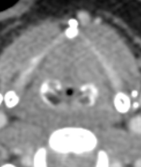
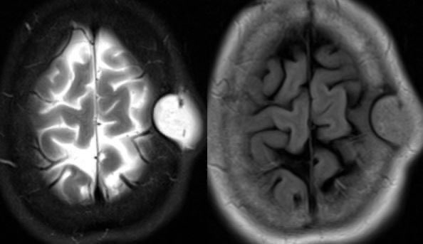
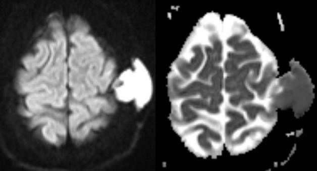
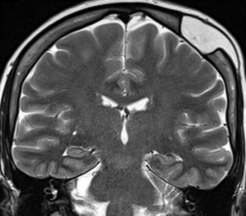
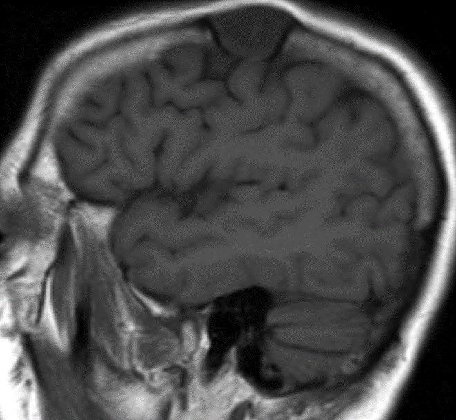
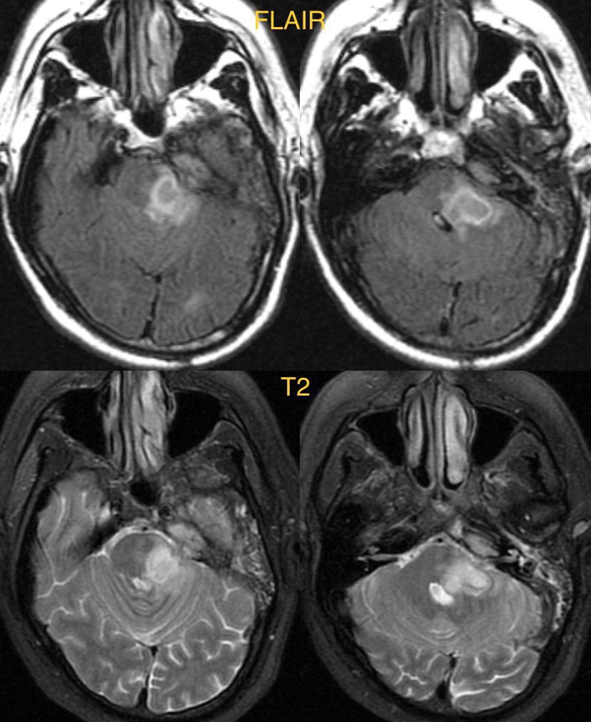
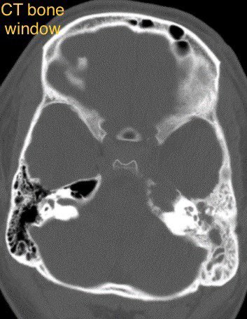
Patient presents with altered mental status. Unenhanced CT shows discrete hypodense foci in the bilateral paramedian thalami
#radres #futureradres #radtwitter @ACRRFS @RSNA Image
Differential diagnosis includes:
Top of the basilar artery syndrome
Artery of Percheron infarct
Bilateral internal cerebral vein thrombosis
Answer: Artery of Percheron infarct

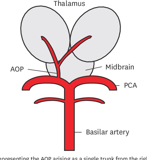
The artery of Percheron is a rare anatomic variant where a single artery supplies both medial thalami, typically arising from the P1 segment of the PCA. Image
Top of the basilar artery syndrome and bilateral internal cerebral vein thrombosis and be excluded by proving patent on CTA. Additionally, thrombosis of the internal cerebral veins tends to create more edema in the bilateral thalami rather than discrete focal infarct.

• • •

Missing some Tweet in this thread? You can try to force a refresh
 

Keep Current with Daniel Gewolb, MD

Daniel Gewolb, MD Profile picture

Stay in touch and get notified when new unrolls are available from this author!

Read all threads

This Thread may be Removed Anytime!

PDF

Twitter may remove this content at anytime! Save it as PDF for later use!

Try unrolling a thread yourself!

how to unroll video
  1. Follow @ThreadReaderApp to mention us!

  2. From a Twitter thread mention us with a keyword "unroll"
@threadreaderapp unroll

Practice here first or read more on our help page!

More from @daniel_gewolb

Mar 23
Neuroimaging checklist for the patient with Temporal Lobe Epilepsy (TLE) 🧠 ✅

Credit to the excellent talk on the @TheASNR fellowship curriculum given by @EMiddlebrooksMD for inspiration on this topic

#MedEd #medicine #radres #Neurology #Neurosurgery #futureradres


Image
Image
Image
Image
1.Encephalomalacia (usually from old infarct, trauma, etc.)
2.Mesial Temporal Sclerosis
3.Sphenoid encephaloceles
4.Prominent arachnoid pits (may be encephalocele precursors)
5.Temporal lobe epilepsy with Amygdala enlargement
6.Focal Cortical Dysplasia
7.Polymicrogyria
8.Gray matter heterotopia
9.Tumors
10.HSV encephalitis
11.Autoimmune encephalitis
12.Hypothalamic hamartoma
13.Other infections (NCC, TB, etc.)
14.Incomplete hippocampal inversion (uncertain significance)
15.Others, please add 🙏
Read 20 tweets
Feb 2
What is the most likely diagnosis in this 70 y/o w/ history of pontine infarct ~8 months ago now presenting with worsening ataxia and dysmetria?

#MedEd #radres #Neurology #neurosurgery #futureradres #Radiology


Image
Image
Image
Image
Answer (probable): Wallerian degeneration of the pontocerebellar fibers

🔷I only have 1 time point w/o follow up nor images of the pontine infarct. This patient also had cirrhosis. It is possible that the tracts have degenerated due to hepatic encephalopathy or other process
🔷Regardless of the cause in this case, it is important to be aware of these fiber tracts and their appearance when degenerated. Additionally, many other diagnoses can look similar and involve the bilateral middle cerebellar peduncles w/ differential in 🧵
Read 14 tweets
Jan 12
What is the most likely diagnosis in this 75 y/o M w/ history of Alzheimer’s disease on lecanemab (last infusion ~1 week prior) presenting w/ headache and confusion? 🧠

More images in 🧵

#MedEd #Neurology #medicine #radres #Neurosurgery #futureradres @a_charidimou


Image
Image
Image
Image
More images 👇

Image
Image
Image
Answer: Amyloid-related imaging abnormalities (ARIA)

🔷ARIA is a term used to describe a spectrum of imaging findings in patients receiving anti-amyloid beta immunotherapies for Alzheimer’s disease

💡 Tips and learning points for this complex topic are welcome as always 🙏
Read 15 tweets
Jan 7
Imaging Quick Tips & Tricks for Cranial Neuropathies 🧠

A neuroimaging 🧵 on check areas and pathology

#neurology #meded #medicine #neurosurgery #ent #radres #futureradres @AlbanyMedRadRes @PennRadiology


Image
Image
Image
Image
💡 General imaging tips:

1️⃣Big issues and certain cranial nerves see on routine MR
2️⃣Thin slice high res axial and coronal T2 sequences (FIESTA, CISS, SPACE, etc.) useful for cisternal segments
3️⃣T1C- and T1C+ w/ fat sat best for extracranial segments
4️⃣CT often complementary
🔷OLFACTORY NERVE (CN 1)

📺 Imaging:
▶️Not well seen on routine brain MRI
▶️Seen well on coronal T2 MR
▶️Include medial temporal lobes in assessment
▶️Nasal vault and cribriform plate seen well on coronal CT of sinuses or orbits
Image
Image
Read 36 tweets
Dec 23, 2023
What is the most likely diagnosis in this 25 y/o M presenting with right facial paralysis and pulsatile tinnitus?

More images in 🧵

#ENT #MedEd #Neurology #Neurosurgery #radres #futureradres @ASHNRSociety


Image
Image
Image
Image
More images 👇


Image
Image
Image
Image
Answer: Temporal Bone facial nerve venous malformation (Hemangioma)

🔷These are benign congenital venous malformations
Read 13 tweets
Nov 19, 2023
Intracranial HYPOtension, imaging findings and diagnostic approach

Imaging and learning points in 🧵 🧠

#Neurology #neurosurgery #MedEd #radres #futureradres #neurorad @The_ASSR


Image
Image
Image
Image
Clinical:

▶️Headache that is worst when upright and improves with lying flat as it gets the sagging brain off the jagged skull base (at least that’s how I remember it)

💡May also present with chronic headaches or without headaches
▶️Rarely can have abducens palsy-> the nerve is anchored in Dorello’s canal so brainstem sagging can stretch the nerve 🧠

▶️Anything that reduces the volume of CSF can lead to intracranial HYPOtension
Read 16 tweets

Did Thread Reader help you today?

Support us! We are indie developers!


This site is made by just two indie developers on a laptop doing marketing, support and development! Read more about the story.

Become a Premium Member ($3/month or $30/year) and get exclusive features!

Become Premium

Don't want to be a Premium member but still want to support us?

Make a small donation by buying us coffee ($5) or help with server cost ($10)

Donate via Paypal

Or Donate anonymously using crypto!

Ethereum

0xfe58350B80634f60Fa6Dc149a72b4DFbc17D341E copy

Bitcoin

3ATGMxNzCUFzxpMCHL5sWSt4DVtS8UqXpi copy

Thank you for your support!

Follow Us!

:(