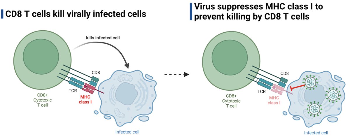
A while ago, @MiyuMoriyama et al showed that SARS-CoV-2 variants suppress MHC I levels in infected cells to the same degree as the ancestral virus. Then came the Omicron variants. A short update. (1/) biorxiv.org/content/10.110…
Since the original submission, @MiyuMoriyama, with the help of @NathanGrubaugh's team & @carolilucas, obtained and analyzed the ability of Omicron subvariants shown here 👇🏽 Miyu gated on spike-positive (infected) cells and compared MHC I levels to uninfected (S-) cells. (2/)
Note that MHC I surface levels are only downregulated in the infected (S+) cells, but not in the uninfected cells (S-) in the same tissue culture wells. SARS-CoV-2 evades recognition of the infected cells by cytotoxic T cells but has no impact on the surrounding cells. (3/)
New data are pretty striking. Compared to the ancestral (WA1) or Epsilon (B.1.429) variant, Omicron subvariants BA.1, BA.2.12.1, XAF and BA.4 all suppressed MHC I surface expression better in infected cells. An irrelevant surface marker (CD324) was not affected by infection. (4/)
Our new data demonstrate the evolution of the Omicron variants in further suppressing MHC I expression and escaping detection/killing by CD8 T cells. Thus, in addition to evasion from Ab and innate immunity, MHC I ⬇️ by Omicron may further ⬆️ replication & transmission. (End)
• • •
Missing some Tweet in this thread? You can try to
force a refresh
Very excited to share our latest research on immunological features of #LongCovid. Our 2+ year collaboration with @PutrinoLab with many other fantastic colleagues and patients - Mount Sinai Yale Long COVID (MY-LC) study by @sneakyvirus1 et al. 🧵(1/)
There are multiple hypotheses behind long COVID pathogenesis including persistent virus/viral remnants, autoimmunity, dysbiosis, virome reactivation and tissue damage. Our data will dive deep into some of these. (3/)
A brilliant & timely review by Prof. #DianeEGriffin on the persistence of viral RNA following RNA virus infection - which can be associated with late progressive disease or nonspecific lingering symptoms of post-acute infection syndromes (#PAIS). (1/)
First question addressed is WHERE viral RNA can persist. After a variety of RNA virus infection, viral RNA can persist not only in immune privileged sites (brain, eyes, and testes), but also in blood, lymphoid tissue, joints, respiratory tract, GI tissues, and kidney. (2/)
Consequences of persistent viral RNA may include organ-specific as well as nonspecific postviral syndromes such as long COVID, post-Ebola, and post-polio syndromes, characterized by symptoms including fatigue, headache, muscle pain, and joint pain. (3/)
With millions of #longCOVID patients, it is becoming better known that even a mild infection can lead to longterm debilitating health problems. SARS-CoV-2 joins the long list of other pathogens that cause post-acute infection syndrome (PAIS). (2/)
What are some common and distinct symptoms associated with PAIS? Strikingly, there are a number of shared symptoms such as excertion intolerance, fatigue, pain, neurological symptoms..etc. Others are more unique to the pathogen that triggered the disease. (3/)
In this study, @MiyuMoriyama et al investigate how well SARS-CoV-2 variants of concern (VOC) suppress MHC I needed for recognition by cytotoxic T cells. This question is important to understand how well the virus limits CD8 killing 🧵(1/) @biorxivpreprint biorxiv.org/content/10.110…
CD8 T cells help fight off viral infection by detecting and killing infected cells. CD8 T cells detect MHC I + viral peptide on infected cells. One of the common tricks viruses use to avoid killing is to inhibit MHC I expression and presentation. (2/)
SARS-CoV-2 is no exception. A previous study showed that SARS-CoV-2 (ancestral) induced MHC I down-regulation in infected cells. They found a key role of ORF8 in this process. (3/)
A Phase 2 clinical trial of oral camostat mesylate during early phase of COVID-19 in outpatients reduced illness course (including fatigue) and prevented loss of smell and taste!
Work of fantastic colleagues at @YaleMed. (1/)
This randomized double-blind placebo-controlled phase 2 trial gave patients (within 3 days of testing PCR+) either oral camostat mesylate or placebo pills, 4x/day for 7 days. Note the lower smell/taste scores (meaning better ability to smell and taste) in camostat group (2/)
Camostat mesylate blocks TMPRSS2, which cleaves the spike protein allowing the virus to fuse with the cell and start to replicate. However, there was no differences in detectable viral RNA levels in patients treated with camostat vs. placebo in nasopharyngeal swab or saliva. (3/)
This new preprint by Stadler et al. integrated data from 37 randomized controlled trials to ask how the timing and dose of passive antibodies (monoclonal Ab & convalescent plasma) predict protection from SARS-CoV-2 disease. A short 🧵 (1/)
Timing: the study found that the earlier the patients were treated with monoclonal antibodies (mAb) or convalescent plasma (CP), the more effective the passive antibodies were in preventing the clinical outcome measured (indicated by right end of line). #TheEarlierTheBetter (2/)
These data are reminiscent of endogenously induced antibody responses against SARS-CoV-2. In patients with fatal COVID, the onset of antiviral antibodies was significantly delayed compared to those who survived COVID. @carolilucas@sneakyvirus1 (3/)