IMCrit Profile picture
Jan 29 24 tweets 14 min read
ICU stories (a brief one): One hour before the end of the am shift, u walk around in the ICU to make sure thinks look OK before u type your sign-out note. You spot the resp therapist & the nurse bagging the pt in Rm 306. From the hallway, u see the monitor: HR 160, RR/45, Sat 70%
This is a 30 yo pt w hx of a catastrophic brain bleed, s/p trach & PEG, admitted 2 wks ago w MDR Klebsiella UTI. Doing well, on trach mask 28%, until the episode of acute/unexpected desaturation
When u examine the pt, s/he is in extremis (accessory muscle use-tachycardic-tachypneic-diaphoretic). BP: 105/55. You grab the stethoscope that the resp therapist wears around his neck & you hear breath sounds in both sides (pt is skinny...)
The pt's lips/skin color looks like your scrubs. Actually, these are my scrubs & I am exaggerating about the color (but not too much...)
You ask the resp therapist to add a PEEP valve to the Ambu bag but O2 sat goes only up to the high 80s-90% & drops again. What is your next step?
In a small community hospital, you don't have an army to help. In these cases, I have a very low threshold of sedating/paralyzing/connecting to the ventilator. That's exactly what we did. This "liberated" the resp therapist & the nurse to do "other stuff"...
Isn't it funny that when a pt drops the O2 sat, we take him/her off the vent? I usually try to put them back or keep them on the vent... 🤷‍♂️ @Thind888 has written a great piece about it. The vent waveforms sometimes can give a lot of info (but did not help in this case...)
Now what? The CXR will take at least 20-30 min. Chest CT may be not the best idea since pt is unstable. It's POCUS time! You start from the anterior L lung:
And move to the anterior R lung:
Where do you see lung sliding?
There is some movement in both sides, right? But, they don't look the same, do they? You want to be fancy & you decide to use M-mode. This is the R lung:
How do we call the sign shown inside the M-mode yellow box?
The horizontal lines are called stratosphere (S) or barcode (BC) sign; they are the same thing. However, they should be BELOW, not above, the pleural line. So, in this case, there is no S/BC sign. But,
under the pleural line, there are some vertical lines that are synchronous with the heartbeats. This is "lung pulse" & suggests that the parietal & visceral pleura are in touch but regional ventilation is impaired.
This 👇 was the normal L lung's M-mode for comparison. There is a "sandy" pattern below the pleural line; it is called "seashore sign":
Since there was no strong evidence of pneumothorax on the R side, we decided to do a bronch w a single use bronchoscope (no COI) & this is what we saw:
Not good...
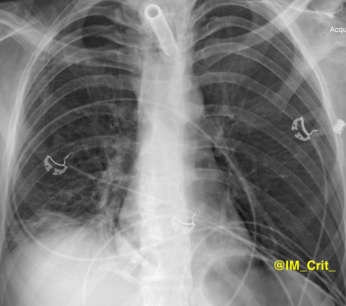
After multiple mucus plugs were suctioned out from all the R lung lobes, oxygenation improved. Post-bronch chest x-ray attached:
Take-home messages:
1. We tend to think that acute severe desaturation is either pneumothorax (tension, if vital signs change dramatically) or PE, but it is not unusual to find atelectasis as the underlying etiology
2. Lung POCUS can be very helpful but be cognizant of the limitations. It can be VERY tricky (I hope @kyliebaker888 agrees with me!)
3. Presence of lung sliding rules out pneumothorax but its absence does not equal w the presence of PTX
4. Lung sliding is taught as an all-or-nothing phenomenon but sometimes there is SOME lung sliding (not complete absence but no presence either) and this suggests decreased regional ventilation (I think!)
5. The seashore & the lung pulse signs are helpful especially in unclear cases (ie, there is no lung sliding but no lung point to confirm PTX) but are not easy especially if you are full of adrenaline & your hand is not steady
Thanks for reading! This approach worked for me in this case but there may be better ways!

#POCUS #ECHOFIRST #POCUSpeeps #FOAMus #FOAMcc #IMPOCUS #MedTwitter #MedEd #EMBound @RJonesSonoEM @jaffa_md @katiewiskar @msiuba @NephroP @MynephCC @HeyDrNik @ICUltrasonica @khaycock2

• • •

Missing some Tweet in this thread? You can try to force a refresh
 

Keep Current with IMCrit

IMCrit Profile picture

Stay in touch and get notified when new unrolls are available from this author!

Read all threads

This Thread may be Removed Anytime!

PDF

Twitter may remove this content at anytime! Save it as PDF for later use!

Try unrolling a thread yourself!

how to unroll video
  1. Follow @ThreadReaderApp to mention us!

  2. From a Twitter thread mention us with a keyword "unroll"
@threadreaderapp unroll

Practice here first or read more on our help page!

More from @IM_Crit_

Jan 15
Judging from yesterday's post, many friends are interested in how to get the most out of these books 👇 and ace the exams, so a few more tips are on their way:
1. Tylenol use can lead to high anion gap metabolic acidosis. Don’t ask me about the mechanism!
2. Every patient who manages to fly eastbound with Southwest Airlines & subsequently develops pneumonia not responding to common antibiotics has actually blastomycosis
3. Every oncology patient who receives chemo is destined to develop tumor lysis syndrome. Please learn about hydration/allopurinol/rasburicase
4. Along these lines, every oncology patient on immunotherapy will develop pneumonitis. Remember the steroids from yesterday’s post?
Read 8 tweets
Jan 15
These books from @accpchest & @SCCM represent my study goal for this month. Before starting any (re-)certification exam & especially f you want to ace the tests, there are a few recent trends (& old habits/tricks) that you need to be aware of:
1. If there is an option of "doing nothing", this is most likely the correct answer.
2. There is always a mixed metabolic disorder. Memorize Winter's formula.
3. Prepare for several COVID-19-related questions. No surprise here...
4. TEG is very popular. Even of you are a dinosaur, you have to learn the basics about visco-elastic tests.
5. I know you have no CAR T-cell therapies in your hospital (& no one can really spell them correctly), but be prepared for managing cytokine release syndrome.
Read 9 tweets
Jan 13
ICU stories: 65 yo pt, fairly healthy besides HTN & an episode of diverticulitis 3 y ago, is brought to the ED due to 2 wks' hx of abd pain & 1 d hx of N/V ("coffee-ground"). Looked "bad". SBP in 60s - improved to 80s w ivf. Intubated. Had CT A/P 👇:
While you review the CT images, you get the lab results: Lactate 10, WBC 3K, INR 2.0, BUN/Creat 100/3.0, CRP 500 mg/l, Procalcitonin 300. The ED is calling you for the admission. What consult(s) do you ask?
The CT A/P showed a large amount of free intra-peritoneal air; stomach & SB were mildly distended & partially fluid-filled. There was colonic diverticulosis without diverticulitis & mild wall thickening involving the descending colon & the sigmoid colon
Read 13 tweets
Jan 12
ICU stories: 70 yo pt without medical hx but tobacco use (2 ppd x 40 y) was admitted w shortness of breath a wk ago. CXR/chest CT without PE/infiltrate. Was in afib/RVR on admission; placed on heparin & dilt/b-blocker (w some hypotension). Remained dyspneic, at times restless,
“requiring” multiple sedatives, & eventually was brought to the ICU. Intubated for "resp distress" & mental status changes. "Formal" echo, the day of ICU transfer, showed “LVEF 20% w global LV dysfunction”. On the vent 50% - peep 10. BP 110-130/60-70. Lactate < 2.0
Cards follow for "well compensated heart failure". A look w POCUS upon ICU admission:
Read 20 tweets
Dec 26, 2022
How do you examine the lower extremity venous system when you look for deep vein thrombosis? What points do you check with the probe? Do you use Doppler? What are the recommended protocols? The Society of Radiologists in Ultrasound recommends a complete duplex ultrasound:
👆 The black rectangles represent the extent of the compression US. The gray rectangles are the sites of Doppler.
2-CUS (2-points compression US) includes compression of the femoral veins 1-2 cm above & below the saphenofemoral junction & the popliteal veins
up to the calf veins
ECUS (extended compression US), includes compression US from the common femoral vein through the popliteal vein up to the calf veins confluence

CCUS (complete compression US), includes compression US from the common femoral vein to the ankle
Read 9 tweets
Dec 23, 2022
Following up on a discussion during ICU rounds this am: Like most laboratory values in medicine, pH and lactate levels should be evaluated in their context. In this 👇 old study of 6 male oarsmen who participated in a maximal effort on a rowing ergometer, Image
the two lowest pH values were 6.74 and 6.76 (corresponding to [H+] of > 180 nmol/L); the HCO3 levels were undetectable. The lowest lactate level was 32 mmol/l. The oarsmen remained conscious and did not require medical help Image
The conclusion of the study was that "in healthy humans, pronounced, but transient,acidosis is well-tolerated". Finding a pH of 7.05 or a lactate of 10 is usually not a big deal in a patient with DKA or (post-)seizures
Read 4 tweets

Did Thread Reader help you today?

Support us! We are indie developers!


This site is made by just two indie developers on a laptop doing marketing, support and development! Read more about the story.

Become a Premium Member ($3/month or $30/year) and get exclusive features!

Become Premium

Don't want to be a Premium member but still want to support us?

Make a small donation by buying us coffee ($5) or help with server cost ($10)

Donate via Paypal

Or Donate anonymously using crypto!

Ethereum

0xfe58350B80634f60Fa6Dc149a72b4DFbc17D341E copy

Bitcoin

3ATGMxNzCUFzxpMCHL5sWSt4DVtS8UqXpi copy

Thank you for your support!

Follow Us on Twitter!

:(