Ali M. Erturk Profile picture
Apr 6 4 tweets 3 min read Twitter logo Read on Twitter
😮~60% of us who had COVID still might have lingering viral spikes in our heads! Our new study reveals SARS-CoV-2 spike accumulation in the skull-meninges-brain axis & its implications in long COVID. By @zhouyi_rong @HongchengM @Sakethkapoor🔬🧠🦠🧵👇 biorxiv.org/content/10.110…
2/n Summary:
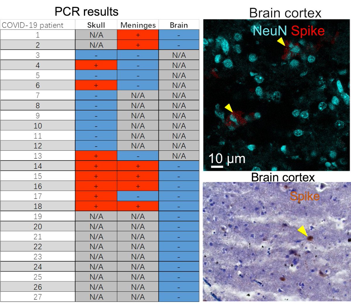
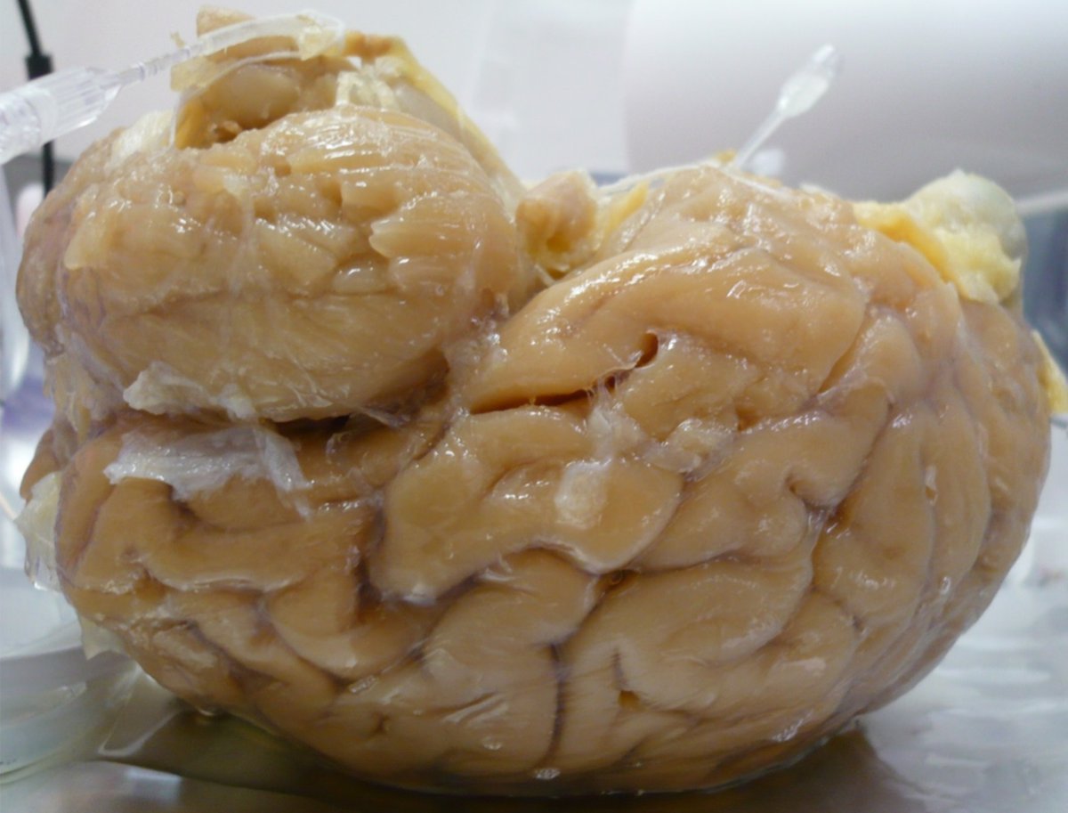
We found SARS-CoV-2 spike protein in the skull-meninges-brain axis in mouse models and human post-mortem tissues long after their COVID, which was associated with vascular and inflammatory changes in the brain along with neuronal damage. Image
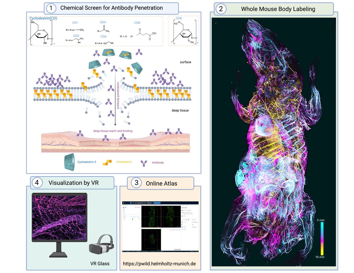
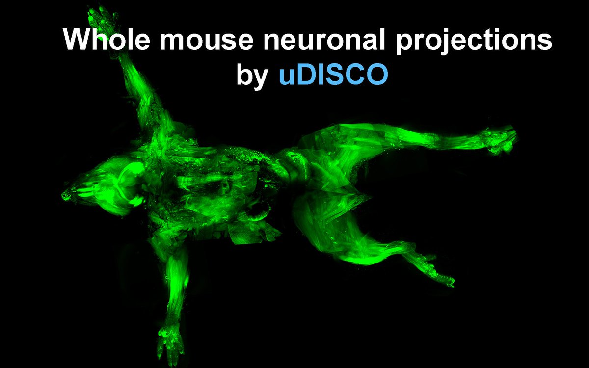
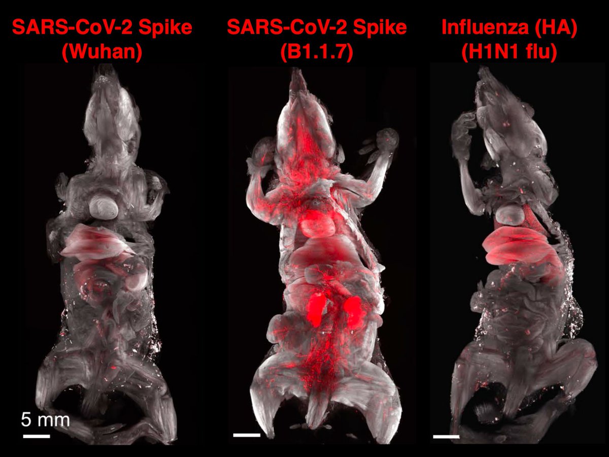
3/n Approach: To discover all tissues that are targeted by SARS-CoV-2, we used unbiased DISCO clearing technology and mapped tissues hit by coronavirus spike vs. Influenza HA proteins (flu). Image
Here is the Tweetorial if you don’t see the rest of tweets:

• • •

Missing some Tweet in this thread? You can try to force a refresh
 

Keep Current with Ali M. Erturk

Ali M. Erturk Profile picture

Stay in touch and get notified when new unrolls are available from this author!

Read all threads

This Thread may be Removed Anytime!

PDF

Twitter may remove this content at anytime! Save it as PDF for later use!

Try unrolling a thread yourself!

how to unroll video
  1. Follow @ThreadReaderApp to mention us!

  2. From a Twitter thread mention us with a keyword "unroll"
@threadreaderapp unroll

Practice here first or read more on our help page!

More from @erturklab

Apr 6
4/n Finding 1)
Along with many organs, we discovered spike accumulations in the skull marrow niches and recently discovered skull-meninges connections (SMCs), revealing a new route of pathogens into the brain. Image
5/n Finding 2)
Critically, we found the spike protein also in the skull bone marrow niches, and meninges of people who died from COVID-19. Image
6/n Finding 3)
Although COVID-19 patients' brain tissue was mostly PCR-negative, spike protein was present in the brain, suggesting a longer half-life compared to viral particles. Image
Read 8 tweets
Feb 20
Imagine you could stain whole adult mice with standard IgG antibodies. What you couldn't do! We present wildDISCO technology for this🤗, creating the first whole body vascular, lymphatic, and nerve atlases. By @HongchengM @jieluo692 biorxiv.org/content/10.110… 🧵👇🏼(turn on the sound)
2/n Summary:
wildDISCO is a new tissue chemistry enabling uniform staining of whole bodies with conventional (150 kDa size) IgG antibodies. It reveals the first whole mouse body cellular & structural maps similar to Allen Brain Atlas only that main them at the whole mouse level. Image
3/n State-of-the-art:
While very powerful, whole mouse body clearing and imaging methods rely on transgenic reporters or small dye labeling, limiting their applications extensively. Thus, new tissue chemistry has been needed to overcome this major bottleneck.
Read 14 tweets
Dec 22, 2022
Spatial-omics goes 3D🤗! Out at Cell @CellCellPress 👉🏼 We developed DISCO-MS with @labs_mann, a spatial proteomics technology for specimens fully imaged in 3D. DISCO-MS is aided by robotics and enables the study of diseases at their early stages. cell.com/cell/fulltext/…🧵👇
Summary:
DISCO-MS is a spatial proteomics technology: it enables proteomics analysis of cleared tissues imaged in 3D. DISCO-MS is aided by AI and robotics and yields proteome similar to fresh or fixed samples.
Have questions? Add them below👇 we will answer📖
DISCO-MS Step 1:
We start by rendering a mouse body or human organ optically transparent using robust clearing methods such as vDISCO or SHANEL. Then, after their cell-level light-sheet microscopy scan, we can visualize fluorescently-labeled structures in 3D as complete.
Read 16 tweets
Feb 13, 2020
We are very excited to share our SHANEL method making the intact human organs transparent, just published at Cell. cell.com/cell/fulltext/… by @shan_heather et al.
See short tweetorial for details:
#clearing #imaging #3D #deeplearning #AI @CellCellPress
Mapping the human brain or other organs is a crucial step forward deciphering how they function in health and disease.
Tissue Clearing methods work well on rodent tissue but haven been poorer on stiff & aged human organs. Making the whole human organs transparent required a new approach.
Read 13 tweets
Dec 31, 2018
I am excited to share our publication of vDISCO technology: a whole mouse body clearing & imaging method nature.com/articles/s4159…. The thread below presents details of vDISCO and its biological findings. @NatureNeuro @nature. Happy New Year all! 1/12
2 years ago, we published uDISCO to clear and image intact adult mouse body with light-sheet microscopy. However, neuronal details were mostly invisible through intact bones, skin and highly autofluorescent muscles. nature.com/articles/nmeth…. 2/12
Now, we developed the vDISCO technology to overcome these problems, and generated a clear whole mouse neuronal projection map of the Thy1-GFPM mouse. 3/12
Read 12 tweets

Did Thread Reader help you today?

Support us! We are indie developers!


This site is made by just two indie developers on a laptop doing marketing, support and development! Read more about the story.

Become a Premium Member ($3/month or $30/year) and get exclusive features!

Become Premium

Don't want to be a Premium member but still want to support us?

Make a small donation by buying us coffee ($5) or help with server cost ($10)

Donate via Paypal

Or Donate anonymously using crypto!

Ethereum

0xfe58350B80634f60Fa6Dc149a72b4DFbc17D341E copy

Bitcoin

3ATGMxNzCUFzxpMCHL5sWSt4DVtS8UqXpi copy

Thank you for your support!

Follow Us on Twitter!

:(