Hey #PathTwitter & #MedTwitter, check out Pedro Pascal transformed into Pedro Pathcal as different stains & methods in histo/cyto/pathology!
Who said #Pathology & #LabMed can't be fun? Embrace your inner histo/cyto/pathologist and celebrate #LabWeek2023! (4/23 - 4/29) 🧬🔬🎭
Who said #Pathology & #LabMed can't be fun? Embrace your inner histo/cyto/pathologist and celebrate #LabWeek2023! (4/23 - 4/29) 🧬🔬🎭

Hematoxylin, fancy name for a natural dye derived from tropical trees logwood and Brazilwood. It binds to acids (ie.DNA & RNA), turning them a lovely shade of blue or purple. It's a daily go-to in pathology labs! Check out PMID: 30001639 for a great read on its history and usage.

Eosin? More like "Oh, so pink!" This synthetic dye binds to all things positive in cells and ECM, highlighting muscles and connective tissues in shades of pink and red. It's a routine go-to in pathology labs along with hematoxylin! #PathMemes #LabLife

Periodic Acid-Schiff, the perfect way to see carbs in action! This stain is used daily to highlight glycogen, mucin, and basement membranes. And it's not just for histology- PAS is also used to detect complex carbs in our food. Bet you never thought a stain could be so versatile!

Gomori Methenamine Silver (GMS), not just for detecting fungi and bacteria, it can also reveal the beauty of basement membranes and elastic fibers! They all will stain brown to black. Thanks to George Gomori's genius invention 🧪🔬👨🏻🔬 #pathology
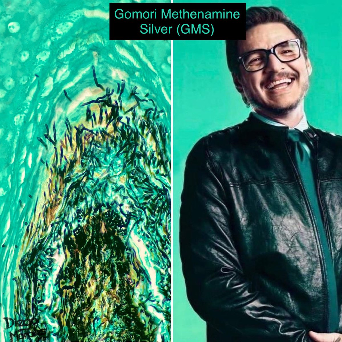
… And here's a fun fact - it also decorates melanosomes black and hair shafts yellow. #PathologyFacts

Alcian blue, a colorful dye that loves acidic mucosubstances like glycoproteins, proteoglycans, and mucins!
Check out my previous tweet about the origin of its name 👉🏼
Check out my previous tweet about the origin of its name 👉🏼
https://twitter.com/diegomoralesn/status/1542334661719908353?s=46&t=VG5l-IOVoh10Pwky1GUcGQ

Masson's trichrome, a three-color stain developed by Claude P Masson (~1929 🇫🇷) that highlights nuclei in blue/black, collagen fibers in blue/green, and cytoplasm and other structures in red. Although it's been reported to show black and green colors, I have yet to see them 🤔

Verhoeff-Van Gieson: The dynamic duo of Fred Verhoeff and Ira T. Van Gieson brought us this stain in 1908🇺🇸. It helps us differentiate elastic fibers from collagen in tissues, with elastic fibers staining black/brown and collagen appearing kind of magenta. 📸 elastic cartilage👂🏼

Fun fact (…a bit of a myth too 🫣): Congo red stain + polarized light = apple green birefringence! 🍏🔍 This phenomenon helps us identify amyloid deposits in tissues. Just another reason why pathology is so cool! #PathTwitter #MedTwitter #PathologyFacts

Got iron? Prussian Blue does! 💙 This nifty stain can detect iron in tissues by producing a stunning blue pigment when it reacts with ferric ions. And the best part? It's highly specific, so it won't stain anything else. How cool is that? 🤩

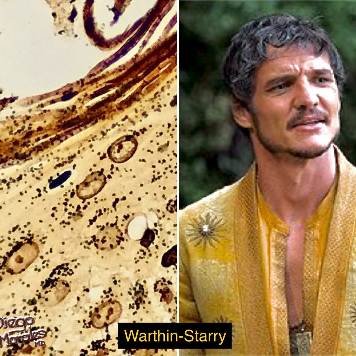
Warthin-Starry, invented by Aldred Warthin and Allen Starry in 1920. Silver-based stain that turns spirochetes black, like the ones causing syphilis and Lyme's. Also handy for finding H. pylori. Not recommended for coloring your hair, though.
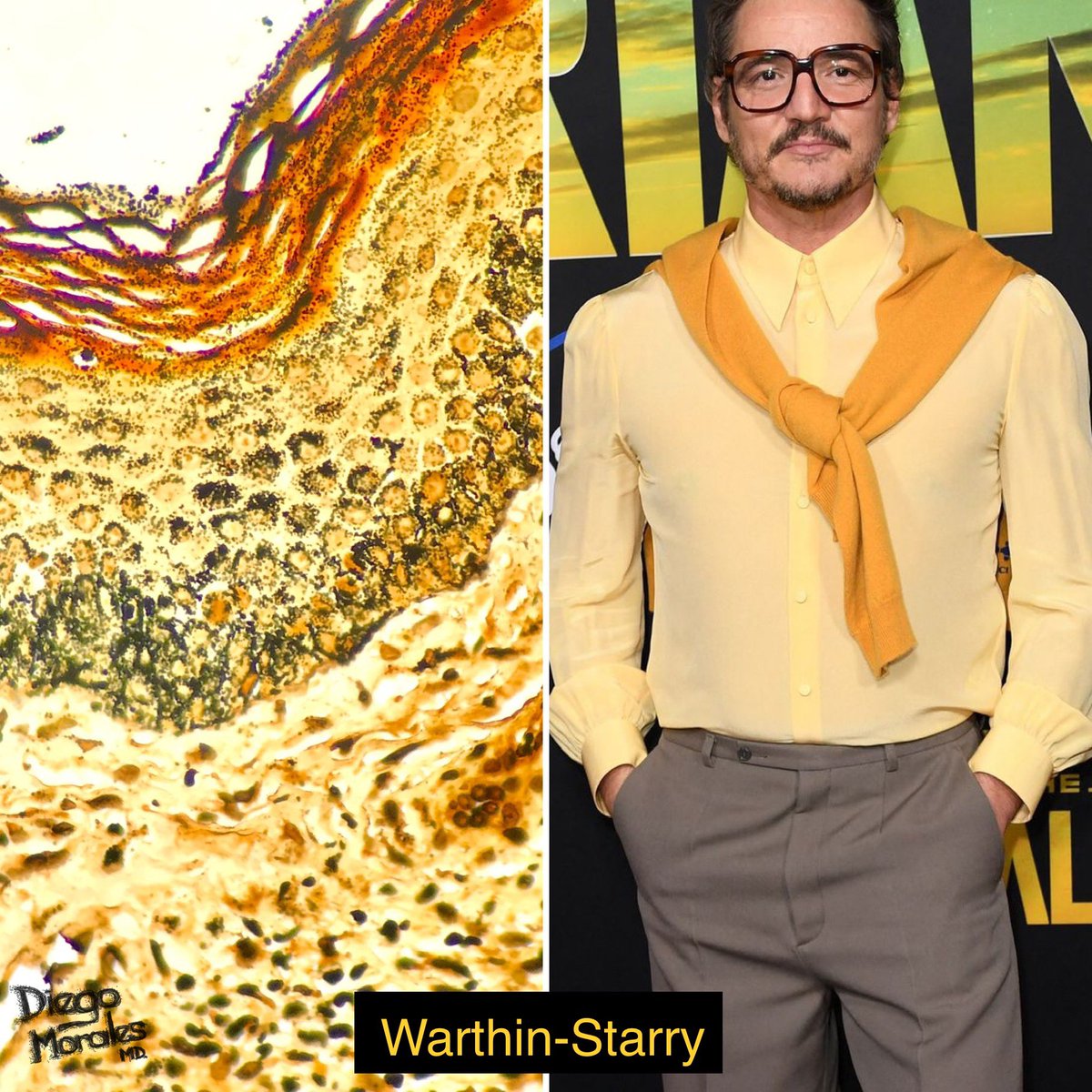
Some people call it "Worthless-Starry"? Harsh. Sure, it's tough to master and has become pretty much obsolete being replaced by T. pallidum #IHC. Buy hey, it brings out the radiance of melanosomes! Cheers for that 🍸🥂
https://twitter.com/diegomoralesn/status/1544630312910692352?s=46&t=xDhgvb0dVeUl15_9nnNLZQ
Colloidal iron: Identifies mucin, despite its name, it doesn’t actually detect Fe, but rather uses a colloidal solution of ferric iron that binds to the mucin molecules, causing them to turn blue, as seen in this granuloma annulare image.

Von-Kossa: Developed by Gustav von Kossa in 1901, this stain detects calcium phosphate crystals in tissues by converting them into a ⚫️ or 🟤 color with silver nitrate and UV light. But be careful, it may miss out on other forms of calcium (i.e. Ca carbonate or Ca oxalate)!

Ziehl-Nieelsen (Acid Fast): It is like a highlighter for Mycobacteria, making them glow under the microscope! Thanks to Franz Ziehl and Friedrich Neelsen, who developed it in 1882, we can now easily spot these tricky bacteria in tissue samples. Very few own a Mycobacteria shirt!

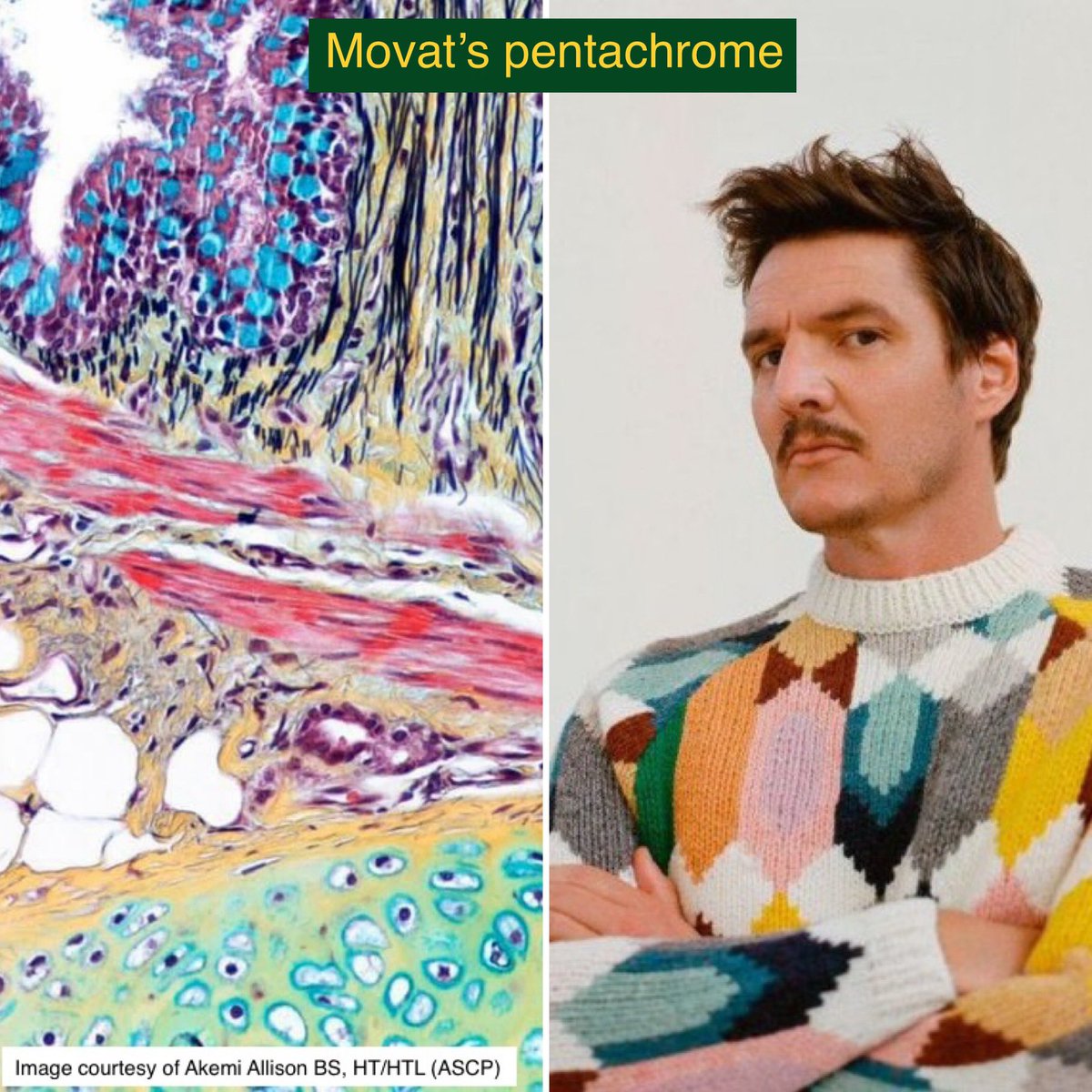
Movat’s pentachrome: Developed by 🇨🇦 Dr. Movat in the 60s:
-black: nuclei & elastic fibers
-blue: mucin
-red: muscle
-yellow: collagen & reticular fibers
-intense red: fibrin
Plus, there's some green magic happening. It might just be the most visually stunning stain out there 🤩
-black: nuclei & elastic fibers
-blue: mucin
-red: muscle
-yellow: collagen & reticular fibers
-intense red: fibrin
Plus, there's some green magic happening. It might just be the most visually stunning stain out there 🤩
Toluidine blue: Basic dye that sticks to nucleic acids, GAGs & other acidic molecules in the EC matrix. Great for visualizing cartilage, mast cells (which stain metachromatically), and other structures rich in acid. During my 🇨🇴 residency, we even used it to detect H. pylori!

Papanicolau: Invented by a Greek-American scientist George Papanicolaou in the 1920s, this simple and cheap test has saved countless lives worldwide by detecting cervical and other types of cancer. A mix of various dyes, it's simply brilliant! 🎉🧬 #PapSmear #CancerDetection

Diaminobenzidine (DAB): The ultimate undercover #IHC agent! It starts off invisible but with the help of horseradish peroxidase (HRP), it turns into a 🟤 precipitate, exposing the antigen's hideout. We use it daily to catch those sneaky antigens in tissues!

Fast Red: #IHC Chromogen + Alkaline Phosphatase = 🔴 precipitate where antigen-antibody bind. I use it for melanocytic lesions, for obvious reasons! 😜

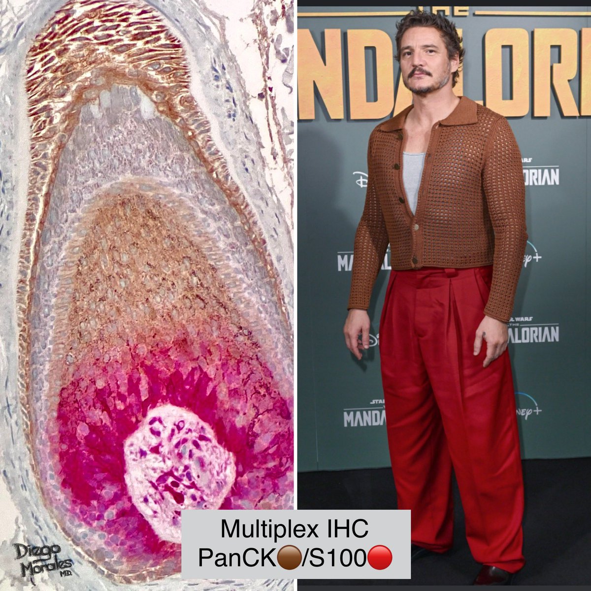
Multiplex #IHC: Double the fun with multiplex IHC! One slide, two antibodies, and a whole lot of color. Check out this hair follicle bulb expressing PanCK (🟤 epithelium) and S100 (🔴melanocytes).
Immunofluorescence, the “mother” of #IHC! With its glowing colors, it's like a party under the microscope. This technique uses fluorescently-labeled antibodies to detect specific proteins in tissues. It's a powerful tool for research and diagnostics. #immunofluorescence

Transmission EM, using heavy metals to create contrast, this technique allows us to visualize and identify different structures in amazing detail. Osmium, uranium, lead, ruthenium are the utilized metals. – Check out 👀 👇🏼this image of the epidermal base!

This Scanning Electron Microscopy (SEM) required deparaffinization and carbon coating to create a stunning 3D effect. And no, those are not baby Yoda's otoliths, but diatoms! SEM is a powerful tool for studying the microscopic world and revealing the beauty hidden in plain sight.

Big thanks to my wonderful wife and involved friends for helping me with “Pathcal project”😅! … but most importantly, let's take a moment during #LabWeek2023 to celebrate all lab workers who make amazing things happen every day. Thank you for your hard work and dedication! 🥳🎊

• • •
Missing some Tweet in this thread? You can try to
force a refresh