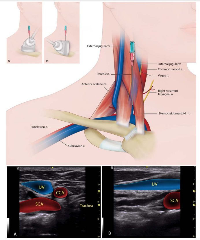
2. The supraclavicular approach to the subclavian vein (A. Transducer in short axis to the vessel, B. long axis) #POCUS#MedEd
3. Infraclavicular approach to the subclavian vein #POCUS#MedEd
4. Femoral vein cannulation (A. short axis #POCUS, B. long axis)
Beware of the anatomic variations of the IJ vein. Use #POCUS if available
Anatomic variations of the subclavian vein. Again, use #POCUS where available.
A: indicates the normal junction and position of the SCV in relation to both the internal jugular vein (IJV) and subclavian artery. B: The SCV may join the IJV as high or even higher than the level of… twitter.com/i/web/status/1…
Finally, thanks to the authors for these great illustrations.
🚨Something to be aware of 👇🏼
Correlate with anatomy above.
#eVExUS (= extended #VExUS) includes evaluation of additional veins when the original 3 veins are unobtainable or unreliable for any reason.
Below👇🧵 is an illustrative case showing abnormal pattern in all these veins.
#POCUS #FOAMed #Nephpearls
1⃣ IVC #ultrasound: dilated
1. A classic #VExUS #POCUS example showcasing how diuretic therapy led to the simultaneous improvement of all three waveforms (hepatic, portal, and intrarenal) alongside improvement in serum creatinine and sodium levels.
🔗 karger.com/bpu/article/51…
2. This example underscores the dynamic nature of the portal vein waveform. Observe the transition from a pulsatile pattern to a continuous one during fluid removal, and its reversal to a pulsatile pattern with positive fluid balance.
🔗 kidneymedicinejournal.org/article/S2590-…
#echofirst views used to evaluate the right heart.
From ASE comprehensive echocardiography textbook/2010 guidelines
#POCUS #MedEd #CriticalCare #IMPOCUS
(🧵of 5 tweets)
Common #echofirst views used in the #POCUS setting, image acquisition pearls and structures visualized.
Thread below🧵
Courtesy, ASE TTE guidelines 2018
#MedEd #FOAMed #FOAMcc
1⃣ Parasternal window
2⃣ Parasternal window continued
3⃣ Parasternal window, short axis plane
Often underutilized, can get an idea of TR when apical window is difficult, can do RVOT Doppler as well
Focused question: Is HTN in a #dialysis patient always⬆️ intravascular volume? Remove💧?
Clinical: ESRD due to polycystic kidney disease, BP 160s/~100, IVC not well-visualized.
Next step to assess RAP: #VExUS
Below is hepatic vein Doppler
#POCUS #Nephpearls #FOAMcc
Follow the🧵 https://t.co/xZe23bHvJVtwitter.com/i/web/status/1…
2/ 👆What do you think?
3/ 👆HV #VExUS is showing resiprophasic D-reversal.
We commonly talk about S-reversal. Where do you see D-reversal? - constrictive pericarditis, tamponade, some cases of severe pulmonary HTN.
Next step: Evaluate the🫀
👇PV looks fine