Lea Alhilali, MD Profile picture
May 26 13 tweets 8 min read Twitter logo Read on Twitter
1/Time is brain! So you don’t have time to struggle w/that stroke alert head CT.
Here’s a #tweetorial to help you with the CT findings in acute stroke.

#medtwitter #FOAMed #FOAMrad #ESOC #medstudent #neurorad #radres #meded #radtwitter #stroke #neurology #neurotwitter Image
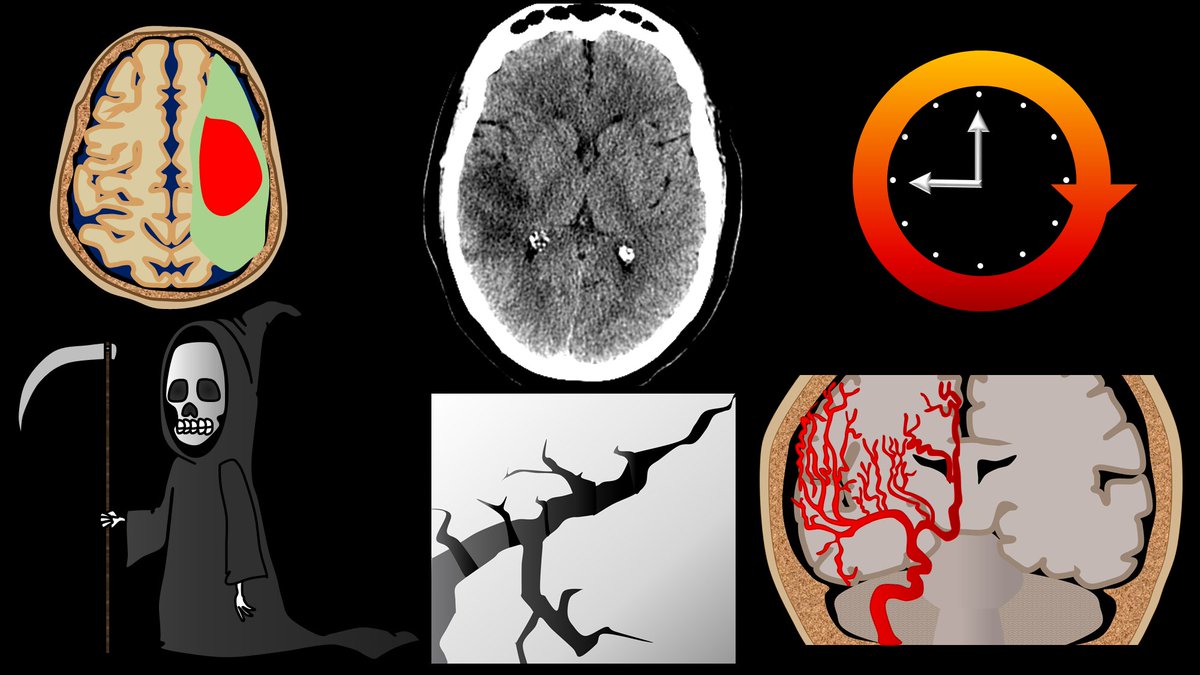
2/CT in acute stroke has 2 main purposes—(1) exclude intracranial hemorrhage (a contraindication to thrombolysis) & (2) exclude other pathologies mimicking acute stroke. However, that doesn’t mean you can’t see other findings that can help you diagnosis a stroke. Image
3/Infarct appearance depends on timing. In first 12 hrs, the most common imaging finding is…a normal head CT. However, in some, you see a hyperdense artery or basal ganglia obscuration. Later in the acute period, you see loss of gray white differentiation & sulcal effacement Image
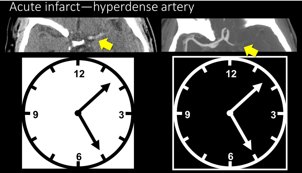
4/Hyperdense artery sign occurs when you see the thrombus in the artery. The thrombus appears hyperdense bc clot is denser than normal flowing blood—& CT is just a measure of density. So an artery filled w/clot will be denser than arteries filled with flowing blood. Image
5/Bc the hyperdensity you are seeing is clot, there will not be flowing blood in this region on CTA. So the hyperdense artery will be the inverse of the CTA--where there is hyperdensity on non-contrast CT, there will be no density/contrast on CTA—like a negative of a photograph Image
6/The other sign in the first 12 hours is the blurred basal ganglia/lentiform nucleus. Usually this region is a triangle of low density white matter (ant limb internal capsule, post limb internal capsule, external capsule) surrounding the high density lentiform nucleus Image
7/In an acute infarct, this triangle becomes blurred, as the lentiform nucleus becomes more edematous, it becomes similar in density to white matter. So instead of clean line between white and gray matter, they look like they are smear together. Image
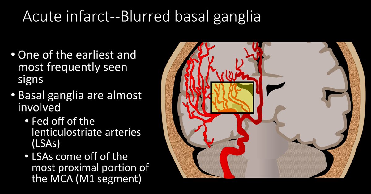
8/The lentiform nucleus is commonly infarcted bc it receives blood from the lenticulostriate arteries that come off of the M1, so unless there is an occlusion more distal in the MCA, the blood supply to the lentiform nucleus is cut off and it infarcts early. Image
9/Why do regions become low density when they infarct? This is bc when O2 & ATP run our, Na/K pump stops working & bc of the osmotic gradient, Na & H20 rush into the cell. More water in the cell = lower density. For every 1% increase in H20 there is a 2.5 HU decrease in density Image
10/This brings us to our next sign—hypodense regions of brain outside the basal ganglia. If the brain is low density, that means it has run out of ATP and swelled, which means the damage is irreversible. Low density = dead brain = poor prognostic sign. Image
11/Another region that infarcts early is the insula. This is bc the insula is actually an internal watershed in the MCA territory. It is the watershed between the lenticulostriates and the M2 sylvian branches, so it will infarct relatively early with low blood supply Image
12/Later you will get sulcal effacement. Normally, the brain should have lots of sulci that look like ice cracks/crevasses along its surface. As more water accumulates in the dead cells, more swelling occurs, and these crevasses become effaced by the swollen brain. Image
13/So now you know the 5 main signs of acute infarct on CT—remember, if you see these five, soon that brain won’t be alive! Image

• • •

Missing some Tweet in this thread? You can try to force a refresh
 

Keep Current with Lea Alhilali, MD

Lea Alhilali, MD Profile picture

Stay in touch and get notified when new unrolls are available from this author!

Read all threads

This Thread may be Removed Anytime!

PDF

Twitter may remove this content at anytime! Save it as PDF for later use!

Try unrolling a thread yourself!

how to unroll video
  1. Follow @ThreadReaderApp to mention us!

  2. From a Twitter thread mention us with a keyword "unroll"
@threadreaderapp unroll

Practice here first or read more on our help page!

More from @teachplaygrub

May 23
1/”Tell me where it hurts.” How back pain radiates can tell you where the lesion is—if you know where to look!

A #tweetorial about how to remember lumbar radicular pain distributions.

#medstudenttwitter #medtwitter #radres #FOAMed #FOAMrad #neurorad #tweetorial #Meded Image
2/Let’s start with L1. L1 radiates to the groin. I remember that b/c the number 1 is, well, um…phallic. So the phallic number 1 radiates to the groin. Image
3/Let’s skip to L3 for a second. I remember L3 is to the knee—easy, it rhymes! Image
Read 8 tweets
May 16
1/Do you want a BASIC approach to skullBASE lesions?

My FINAL tweetorial on skullbase lesions—posterior skullbase & overall approach!

This #tweetorial will teach you to diagnose skullbase lesions by answering only TWO simple questions!

#medtwitter #meded #neurosurgery #radres Image
2/Remember, you can think of pathology at the skullbase like bad things that can happen while running. Bad things can get you from below—like falling into a pothole. They can come from within—like a sudden heart attack, or bad things can strike from above, like a lightning bolt Image
3/Same thing w/the skullbase—bad things can come from below, within, or above. Lesions from below are potholes tripping you up. Lesions from w/in the skullbase are like heart attacks strikning from inside. Lesions from above are the lightning, hitting the skullbase from above Image
Read 14 tweets
May 12
1/Talk about the bases being loaded!

Central skull base has some of the most complicated anatomy & pathology in neuro

Do you know how to approach it?

Here’s a #tweetorial to show you how diagnose lesions at the central skullbase!

#meded #medtwitter #FOAMed #neurosurgery Image
2/Think of the skullbase divisions like different countries—each w/their own culture. Each division has lesions that are specific to it—just like countries have food that are specific to them.

I think the central skullbase looks like Italy, hanging down from the ant. skullbase Image
3/Lesions can involve the central skullbase from below, within, or above

Let’s start from below. Nasopharynx is below the central skullbase. Nasopharyngeal carcinomas (NPC) can invade from below

Using our Italy theme, you can remember this bc NPC look like an Italian meatball Image
Read 21 tweets
May 9
1/It’s called the skullBASE but it’s anything but BASIC!

Does the sight of a skullbase lesion strike fear into your heart?

Never fear! Here’s a #tweetorial about a simple approach to these lesions that will change how you look at these cases

#medtwitter #meded #neurosurgery Image
2/Everyone fears the skullbase. It is so complex that not even experts can agree on a classification for the anatomy.

But you don’t need to know detailed anatomy to be able to give a differential diagnosis for a skullbase lesion that accurate & almost as importantly—short. Image
3/The skullbase is incredibly important. If you think of your brain as master or God of your body, then the skullbase is where the finger of God breathes life into the rest of you. All of the neuronal information from the brain travels through the skullbase to bring you to life Image
Read 23 tweets
May 1
1/Do radiologists sound like they are speaking a different language when they talk about MRI? T1 shortening what? T2 prolongation who?

Here’s a translation w/a #tweetorial introduction to MRI.

#medtwitter #FOAMed #FOAMrad #medstudent #neurorad #radres #ASNR23 #neurosurgery Image
2/When it comes to bread and butter neuroimaging—MRI is definitely the butter. Butter makes everything taste better and packs a lot of calories. MRI can add so much information to a case Image
3/In fact, if CT is a looking glass into the brain—MRI is a microscope. It can tell us so much more about the brain and pathology that affects the brain.

So let’s talk about the basic sequences that make up an MRI and what they can show us. Image
Read 22 tweets
Apr 21
1/Don’t let all your effort be in VEIN!

Developmental venous anomalies (DVAs) are often thought incidental but ignore them at your own risk!

A #tweetorial about how to know when DVAs are the most important finding

#meded #medtwitter #neurorad #neurotwitter #radtwitter #radres Image
2/DVAs aren’t hard to recognize on imaging—they have a typical “caput medusae” appearance.

Dilated medullary veins look like snakes all converging into the medusa head of a large draining vein. Image
3/DVAs are incredibly common—1 in 50 may have one

Although >90% are asymptomatic, that would still results in a relatively large number of patients w/symptoms

Also, w/increasing knowledge of DVA physiology, we may find they’re responsible for more symptoms than we realize Image
Read 21 tweets

Did Thread Reader help you today?

Support us! We are indie developers!


This site is made by just two indie developers on a laptop doing marketing, support and development! Read more about the story.

Become a Premium Member ($3/month or $30/year) and get exclusive features!

Become Premium

Don't want to be a Premium member but still want to support us?

Make a small donation by buying us coffee ($5) or help with server cost ($10)

Donate via Paypal

Or Donate anonymously using crypto!

Ethereum

0xfe58350B80634f60Fa6Dc149a72b4DFbc17D341E copy

Bitcoin

3ATGMxNzCUFzxpMCHL5sWSt4DVtS8UqXpi copy

Thank you for your support!

Follow Us on Twitter!

:(