Acute Pericarditis:
- Inflammation of the pericardium
- May be caused by number of factors: viral/bacterial infection, metastatic tumors, collagen vascular diseases, MI, cardiac surgery, and uremia
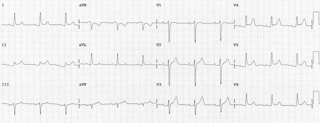
ECG Changes w/ Acute Pericarditis:
- Early phase is characterized by ST segment elevation, due to inflammation of the epicardium, which accompanies inflammation of the overlying pericardium
- Can have generalized ST-T changes in both anterior and inferior leads
Hyperkalemia:
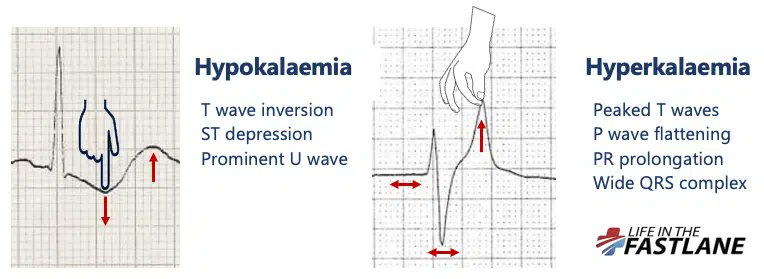
- Distinctive sequence of ECG changes affecting both depolarization (QRS) and repolarization (ST-T)
- First change: Narrowing and peaking of T-waves ('tented' or 'pinched' shape) and can become tall
Hyperkalemia:
- Further elevation: PR intervals become prolonged, P-waves may disappear. Will have intra-ventricular conduction delay, with widening of QRS complexes.
- Can lead to large, undulating (sine wave) pattern with asystole and cardiac death
Inferior Wall Infarction:
- Diaphragmatic portion of the LV
- Will see changes in leads II, III, and aVF
- May produce abnormal Q-waves in these leads
- Generally caused by occlusion of the RCA; less commonly can occur with a left circumflex coronary obstruction
Posterior Infarction:
- Occurs on the posterior (back) surface of the LV
- May be difficult to diagnose because characteristic abnormal ST elevations may no appear in any of the 12 conventional leads
- Tall R-waves and ST depressions can occur in V1 and V2