Lea Alhilali, MD Profile picture
Jun 12, 2023 18 tweets 9 min read Read on X
1/Does PTERYGOPALATINE FOSSA anatomy feel as confusing as its spelling? Does it seem to have as many openings as letters in its name?

Let this #tweetorial on PPF #anatomy help you out

#meded #medtwitter #FOAMed #FOAMrad #neurosurgery #neurology #neurorad #neurotwitter #radres Image
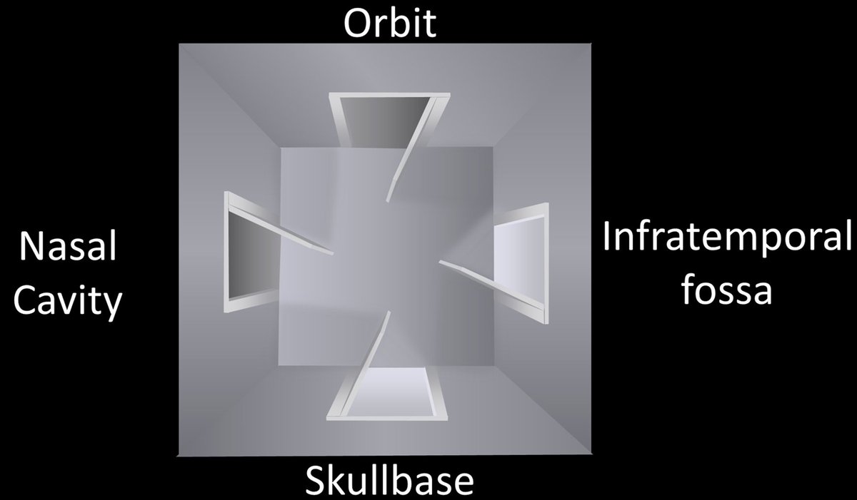
2/The PPF is a crossroads between the skullbase & the extracranial head and neck. There are 4 main regions that meet here. The skullbase itself posteriorly, the nasal cavity medially, the infratemporal fossa laterally, and the orbit anteriorly. Image
3/At its most basic, you can think of the PPF as a room with 4 doors opening to each of these regions: one posteriorly to the skullbase, one medially to the nasal cavity, one laterally to the infratemporal fossa, and one anteriorly to the orbit Image
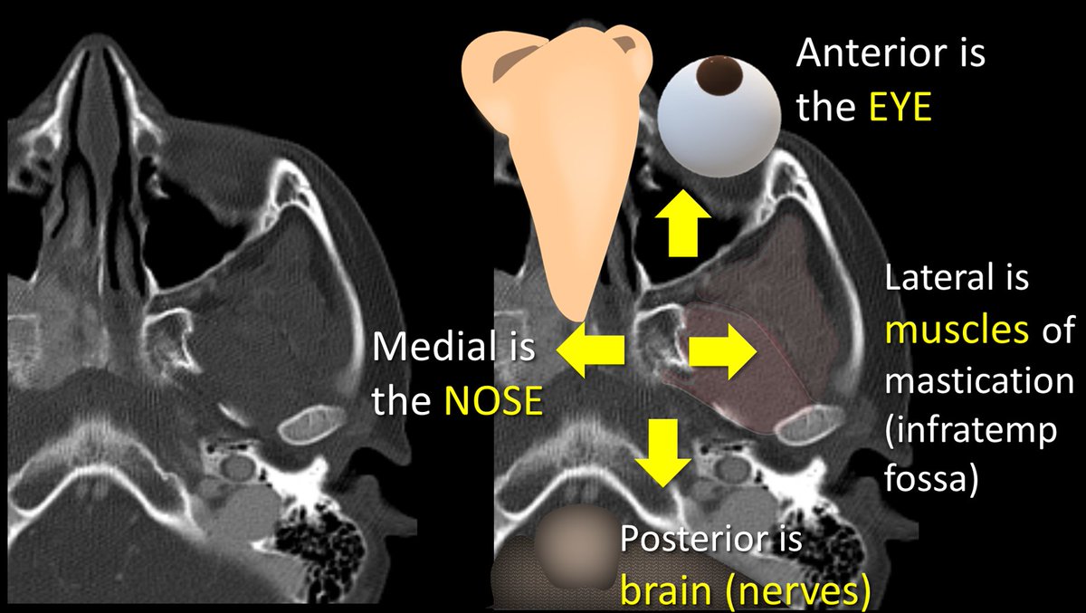
4/You can intuitively remember where each of these doors lead by thinking about what lies around the PPF. Brain is posterior, so post door opens to cranial nerves. Medially is the nose, so med door opens to nasal cavity. Anteriorly is the eye, so ant door goes to orbit & so forth Image
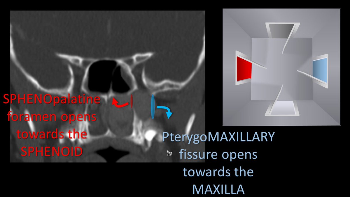
5/Name of each door/opening also tells where they go. SPHENOpalatine foramen goes medially towards the SPHENOID sinus. Inferior ORBITAL fissure is to the ORBIT. PterygoMAXILLARY fissure goes laterally like the MAXILLARY sinus. ROTUNDUM is for a cranial nerve, so goes to the brain Image
6/Let’s start w/the medial & lat openings. Sphenopalatine foramen (SPF) & pterygomaxillary fissure (PMF) can be seen when the walls of the post max sinus & pterygoid plate are parallel. They look like the two round openings of a cylinder—with the cylinder being the PPF itself. Image
7/Their names tell you which side of the cylinder they are on. SPF opens to the sphenoid body/sinus, so it's the medial opening, since these structures are medial. PMF opens towards the maxillary sinus, which goes out laterally towards the zygoma—so PMF is the lateral opening Image
8/There is also an easy mnemonic. In old naval times, the Port side of the ship was the side that docked along the port, so it was the side that dealt w/the outside world. PMF starts w/P, so it is the port side, the side that is towards the world outside the patient (lateral). Image
9/Posterior door has 2 parts. More superior part is foramen rotundum. It looks like a cylinder going straight back. I remember Rotundum is the post door bc both Rotundum & Rear start w/R. You can also remember that Rears are Round, if you want to be a little cheeky—literally Image
10/The lower half of the posterior door is the pterygoid or vidian canal. I remember that Vidian is the lower opening in the posterior door bc the V almost looks like an arrow pointing down. Image
11/Unlike rotundum that looks like a straight cylinder, Vidian has a bit of a curve to it, looking a little bit like the letter L. This helps me to remember that Vidian connects to Foramen Lacerum, bc Vidian looks a little like an L & Lacerum starts w/an L. Image
12/Anterior door is the inferior orbital fissure (IOF). You can remember this bc the eye is anterior to the PPF. You can also remember its name (IOF) with the little mnemonic that “I (eye) Opening is Forward.” Image
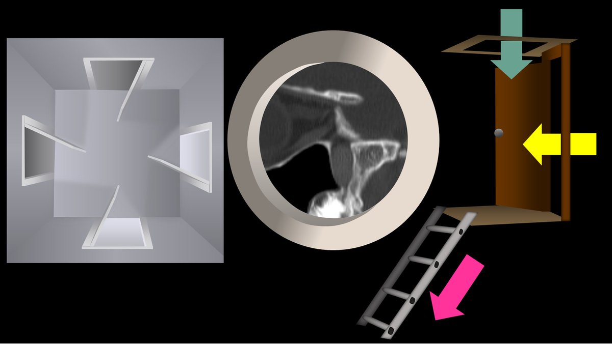
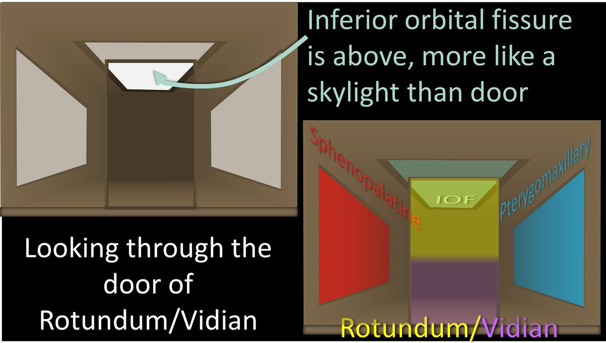
13/But it’s a little bit more complicated than that. While the IOF is anterior, it also superior, more like an anterior skylight than a door. So if you look through the rotundum/vidian door, you will see the SPF medially, the PMF laterally & the IOF in front of you on the ceiling Image
14/But it’s even a little more complicated than that! Turns out there’s a trap door in the floor as well—the palatine canals. You can remember this bc the mouth/palate is below the PPF so you need a hole going down. You can also remember this b/c both Palatine & Pothole start w/P Image
15/So looking from above, you will the Rotundum/Vidian rear door, w/the SPF door medially & the PMF door laterally. Up on the anterior ceiling is the IOF skylight, and down on the floor is the trapdoor to the palatine canals. Image
16/Here is a view from the side. You can better see how the IOF is like a skylight, rotundum is a rear door, and the palatine canals are the trap door with a ladder leading down to the mouth. Image
17/So let’s pretend we are intracranial & peering through the round hole of foramen rotundum. We will see what looks like a loft w/a ladder. The ladder is the palatine canals leading down. The loft itself has a skylight (IOF) & 2 windows—medial window is the SPF & lateral is PMF. Image
18/So now you know the basic anatomy of the PPF. As Elizabeth Taylor once said, “There are many doors in this world, don’t be afraid to look through them.” Don’t be afraid of the doors of the PPF—they can open the world of anatomy to you! Image

• • •

Missing some Tweet in this thread? You can try to force a refresh
 

Keep Current with Lea Alhilali, MD

Lea Alhilali, MD Profile picture

Stay in touch and get notified when new unrolls are available from this author!

Read all threads

This Thread may be Removed Anytime!

PDF

Twitter may remove this content at anytime! Save it as PDF for later use!

Try unrolling a thread yourself!

how to unroll video
  1. Follow @ThreadReaderApp to mention us!

  2. From a Twitter thread mention us with a keyword "unroll"
@threadreaderapp unroll

Practice here first or read more on our help page!

More from @teachplaygrub

Jun 9
1/Need help reading spine imaging? I’ve got your back!

It’s as easy as ABC!

A thread about an easy mnemonic you can use on every single spine study you see to increase your speed & make sure you never miss a thing! Image
2/A is for alignment

Look for:
(1) Unstable injuries

(2) Malalignment that causes early degenerative change. Abnormal motion causes spinal elements to abnormally move against each other, like grinding teeth wears down teeth—this wears down the spine Image
3/B is for bones.

On CT, the most important thing to look for w/bones is fractures. You may see focal bony lesions, but you may not

On MR, it is the opposite—you can see marrow lesions easily but you may or may not see edema associated w/fractures if the fracture is subtle Image
Read 11 tweets
Jun 6
1/Raise your hand if you’re confused by the BRACHIAL PLEXUS!

I could never seem to remember or understand it—but now I do & I’ll show you how!

A thread so you will never fear brachial plexus anatomy again! Image
2/Everyone has a mnemonic to remember brachial plexus anatomy.

I’m a radiologist, so I remember one about Rad Techs.

But just remembering the names & their order isn’t enough.

That is just the starting point--let’s really understand it Image
3/From the mnemonic, we start with the roots—the cervical nerve roots.

I remember which roots make up the brachial plexus by remembering that it supplies the hand.

You have 5 fingers on your hand so we start with C5 & we take 5 nerve roots (C5-T1). Image
Read 20 tweets
Jun 4
1/Having trouble remembering what to look for in vascular dementia on imaging?

Almost everyone w/memory loss has infarcts. Which are important?

The latest @theajnr SCANtastic has what you need to know:

ajnr.org/content/46/5/1…Image
@TheAJNR 2/Vascular cognitive impairment, or its most serious form, vascular dementia, used to be called multi-infarct dementia.

It was thought dementia directly resulted from brain volume loss from infarcts, w/the thought that 50-100cc of infarcted related volume loss caused dementia Image
@TheAJNR 3/But that’s now outdated. We now know vascular dementia results from diverse pathologies that all share a common vascular origin.

It’s possible to lose little volume from infarct & still result in dementia.

So if infarcts are common—which contribute to vascular dementia? Image
Read 20 tweets
Jun 2
1/Having trouble remembering how to differentiate dementias on imaging?

Is looking at dementia PET scans one of your PET peeves?

Here’s a thread to show you how to remember the imaging findings in dementia & never forget! Image
2/The most common functional imaging used in dementia is FDG PET. And the most common dementia is Alzheimer’s disease (AD).

On PET, AD demonstrates a typical Nike swoosh pattern—with decreased metabolism in the parietal & temporal regions Image
3/The swoosh rapidly tapers anteriorly—& so does hypometabolism in AD in the temporal lobe. It usually spares the anterior temporal poles.

So in AD look for a rapidly tapering Nike swoosh, w/hypometabolism in the parietal/temporal regions—sparing the anterior temporal pole Image
Read 16 tweets
May 27
1/Feel perplexed by the lumbosacral plexus??

This plexus doesn’t have to be so complex-us

Here’s what you need to know from this month’s @Radiographics!


@cookyscan1 @RadG_editor doi.org/10.1148/rg.240…Image
@RadioGraphics @cookyscan1 @RadG_Editor 2/The lumbosacral plexus is like a love story

The lumbar & sacral plexuses met & fell in love

They loved each other so much they came together to create the nerves to the lower extremities! Image
@RadioGraphics @cookyscan1 @RadG_Editor 3/Lumbosacral plexus is essentially formed by the nerves from L1-S4 (with some other small contributions)

Remember this bc the plexus is to the lower extremitieis and L & 1 look legs and S & 4 look like feet! Image
Read 12 tweets
May 6
1/Have disagreements between radiologists on the degree of cervical canal stenosis become a pain in the neck?

Worried about sticking your neck out & calling severe cervical stenosis?

This month’s @theAJNR SCANtastic has the latest about Cspine MRI!

ajnr.org/content/46/4/7…Image
@TheAJNR 2/In the lumbar spine, it is all about the degree of canal narrowing & room for nerve roots.

In the cervical spine, we have another factor to think about—the cord.

Cord integrity is key. No matter the degree of stenosis, if the cord isn’t happy, the patient won’t be either Image
@TheAJNR 3/Cord flattening, even w/o canal stenosis, can cause myelopathy.

No one is quite sure why.

Some say it’s b/c mass effect on static imaging may be much worse dynamically, some say repetitive microtrauma, & some say micro-ischemia from compression of perforators Image
Read 16 tweets

Did Thread Reader help you today?

Support us! We are indie developers!


This site is made by just two indie developers on a laptop doing marketing, support and development! Read more about the story.

Become a Premium Member ($3/month or $30/year) and get exclusive features!

Become Premium

Don't want to be a Premium member but still want to support us?

Make a small donation by buying us coffee ($5) or help with server cost ($10)

Donate via Paypal

Or Donate anonymously using crypto!

Ethereum

0xfe58350B80634f60Fa6Dc149a72b4DFbc17D341E copy

Bitcoin

3ATGMxNzCUFzxpMCHL5sWSt4DVtS8UqXpi copy

Thank you for your support!

Follow Us!

:(