Lea Alhilali, MD Profile picture
Aug 10 11 tweets 4 min read Twitter logo Read on Twitter
1/”That’s a ninja turtle looking at me!” I exclaimed. My fellow rolled his eyes, “Why do I feel I’m going to see this on twitter…”

He was right!

A thread about 1 of my favorite imaging findings & pathology behind it

#medtwitter #FOAMed #FOAMrad #meded #neurotwitter #radres Image
2/Now the ninja turtle isn’t an actual sign—yet! But I am hoping to make it go viral as one.

To understand what this ninja turtle is, you have to know the anatomy.

I have always thought the medulla looks like a 3 leaf clover in this region. Image
3/ The most medial bump of the clover is the medullary pyramid (motor fibers).

Next to it is the inferior olivary nucleus (ION).

Finally, the last largest leaf is the inferior cerebellar peduncle.

Now you can see that the ninja turtle eyes correspond to the ION. Image
4/But why are IONs large & bright in our ninja turtle? This is hypertrophic olivary degeneration. It is how ION degenerates when input to it is disrupted.

Input to ION comes from a circuit called the triangle of Guillain & Mollaret—which sounds like a fine French wine label! Image
5/At its simplest, the triangle consists of the ipsilateral red nucleus, ION itself, & contralateral dentate nucleus.

Red nucleus signals the ipsilateral ION, who then send signals to the contralateral dentate, which signals back to the red nucleus & the triangle is complete! Image
6/Signals from the red nucleus to ION are inhibitory.

I remember this bc red=communism=stopping you from doing what you want

So when you disrupt the circuit, the ION is finally gets the green light to crazy & hypertrophies—that’s how you get hypertrophic olivary degeneration! Image
7/The triangle is actually a bit more complex—it also includes the structures that carry the signal between the three points.

So any damage to any of the points of the triangles or the structures connecting them will result in hypertrophic olivary degeneration. Image
8/You get a different appearance depending on where you disrupt the circuit.

If you disrupt it in the brainstem (red nucleus, central tegmental tract), the olivary degeneration will be on the SAME SIDE.

I remember that bc Stem and Same both start with S. Image
9/If you disrupt it in the cerebellum (dentate), you will get CONTRALATERAL degeneration.

I remember this bc Cerebellum and Contralateral both start with C. Image
10/Finally, if you interrupt both limbs (ie get both the superior cerebellar peduncle and central tegmental tract as in this example) you will get bilateral hypertrophic olivary degeneration and our famous ninja turtle!

I remember Both and Bilateral start w/B Image
11/So now you know about hypertrophic olivary degeneration and how different insults cause different appearances.

Hopefully you will remember my ninja turtle sign and spread it around so it truly becomes the official sign of bilateral hypertrophic olivary degeneration!

• • •

Missing some Tweet in this thread? You can try to force a refresh
 

Keep Current with Lea Alhilali, MD

Lea Alhilali, MD Profile picture

Stay in touch and get notified when new unrolls are available from this author!

Read all threads

This Thread may be Removed Anytime!

PDF

Twitter may remove this content at anytime! Save it as PDF for later use!

Try unrolling a thread yourself!

how to unroll video
  1. Follow @ThreadReaderApp to mention us!

  2. From a Twitter thread mention us with a keyword "unroll"
@threadreaderapp unroll

Practice here first or read more on our help page!

More from @teachplaygrub

Aug 2
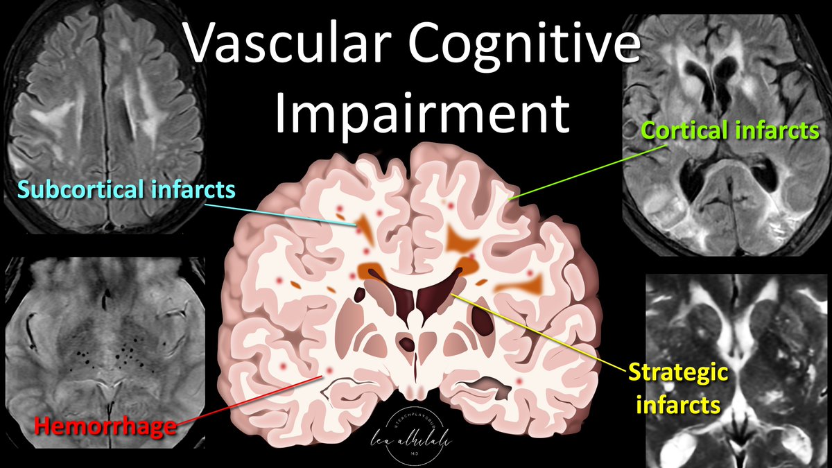
1/Having trouble remembering what you should look for in vascular dementia on imaging?

Almost everyone worked up for #dementia has infarcts. Which ones are important?

Here’s THE FULL #tweetorial this time on the key findings in vascular dementia
#meded #medtwitter #neurotwitter Image
2/Vascular cognitive impairment, or its most serious form, vascular dementia, used to be called multi-infarct dementia.

It was thought dementia directly resulted from brain volume loss from infarcts, w/the thought that 50-100cc of infarcted related volume loss caused dementia Image
3/But that’s now outdated. We now know vascular dementia results from diverse pathologies that all share a common vascular origin.

It’s possible to lose little volume from infarct & still result in dementia.

So if infarcts are common—which contribute to vascular dementia? Image
Read 21 tweets
Jul 21
1/Correlate clinically!

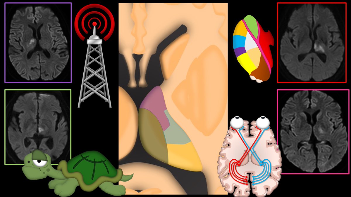
It’s harder than you think in the THALAMUS—where its size is small & but the clinical symptoms are large.

Here’s a #tweetorial to help you remember the main thalamic syndromes & their locations!

#meded #medtwitter #neurotwitter #stroke #radres #FOAMed Image
2/Thalamus is a dense network of nuclei & tracts connected to almost everything in the brain. So almost any symptom can be correlated to it.

So saying “thalamus” as the answer when asked where a lesion is located is always reasonable—even w/o knowing what the symptoms are! Image
3/Think of the thalamus like the internet service provider or ISP for the brain. Like an ISP, everywhere is connected through it.

And like an ISP, things go bad when it goes down.

But just like an ISP, the problems created depend on where in the network the outage is located Image
Read 23 tweets
Jun 30
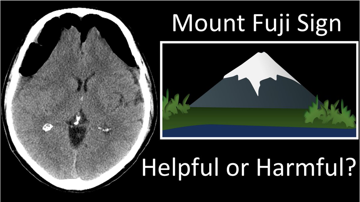
1/Controversy in radiology can get tense! The Mt Fuji sign for tension pnemocephalus is under scrutiny. When should you call it?

A #tweetorial about imaging this important #neurosurgery complication
#medtwitter #FOAMed #FOAMrad #medstudent #radres #meded #neurotwitter
2/Some believe that peaked, mountain like appearance of the frontal lobes is a critical sign of a life-threatening complication & should be called & reported. Others believe it is too non-specific, commonly seen when there isn’t tension & should be retired. Who’s right?
3/First, let’s clarify about what the Mt Fuji sign actually is. Most are familiar with the fact that large collections of pneumocephalus can compress the frontal lobes—making them look like the slopes of a mountain. But this isn’t actually enough to call Mt Fuji.
Read 12 tweets
Jun 23
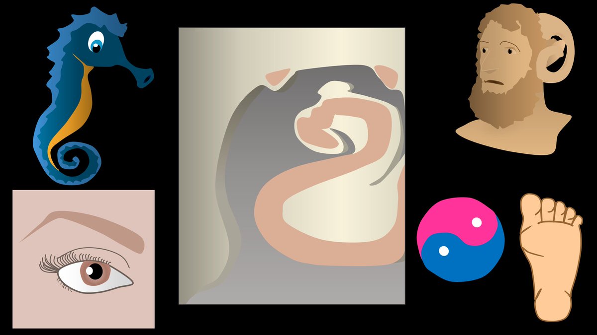
1/ If only there was a way to make hippocampal anatomy memorable!

Here is a #tweetorial of the basics of hippocampal #anatomy that will hopefully stay in your #hippocampus!

#medtwitter #FOAMed #FOAMrad #medstudent #neurorad #radres #neurosurgery #epilepsy #neurotwitter
2/Its name “hippocampus” comes from its shape on gross anatomy.

Early anatomists thought it looked like an upside down seahorse—w/its curved tail resembling the tail of a seahorse.

Hippocampus literally means seahorse.
3/In cross section, it has a spiral appearance, leading to its other name, Cornu Ammonis, translated Ammon’s Horn.

Ammon was an Egyptian god w/spiraling rams horns.

The hippocampal subfields are abbreviated CA-1, CA-2, etc, w/CA standing for “Cornu Ammonis”
Read 14 tweets
Jun 12
1/Does PTERYGOPALATINE FOSSA anatomy feel as confusing as its spelling? Does it seem to have as many openings as letters in its name?

Let this #tweetorial on PPF #anatomy help you out

#meded #medtwitter #FOAMed #FOAMrad #neurosurgery #neurology #neurorad #neurotwitter #radres Image
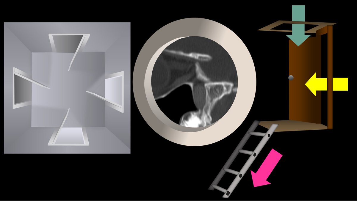
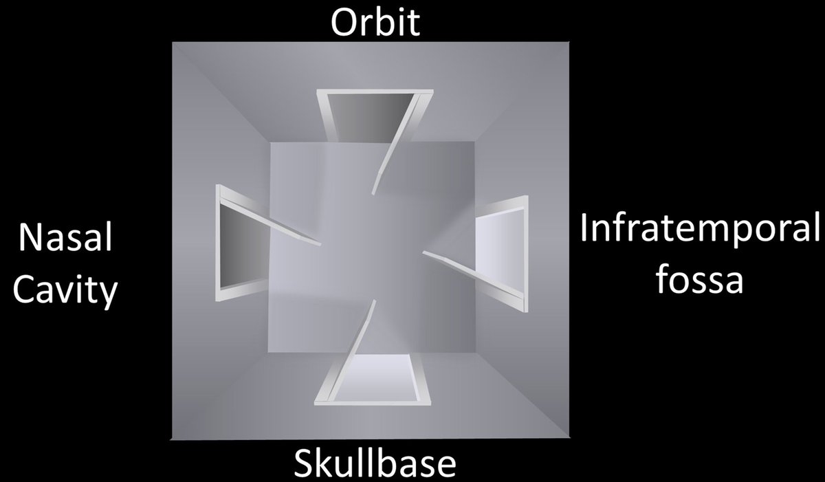
2/The PPF is a crossroads between the skullbase & the extracranial head and neck. There are 4 main regions that meet here. The skullbase itself posteriorly, the nasal cavity medially, the infratemporal fossa laterally, and the orbit anteriorly. Image
3/At its most basic, you can think of the PPF as a room with 4 doors opening to each of these regions: one posteriorly to the skullbase, one medially to the nasal cavity, one laterally to the infratemporal fossa, and one anteriorly to the orbit Image
Read 18 tweets
Jun 9
1/Remembering spinal fracture classifications is back breaking work!

A #tweetorial to help your remember the scoring system for thoracic & lumbar fractures—“TLICS” to the cool kids!

#medtwitter #radres #FOAMed #FOAMrad #neurorad #Meded #backpain #spine #Neurosurgery Image
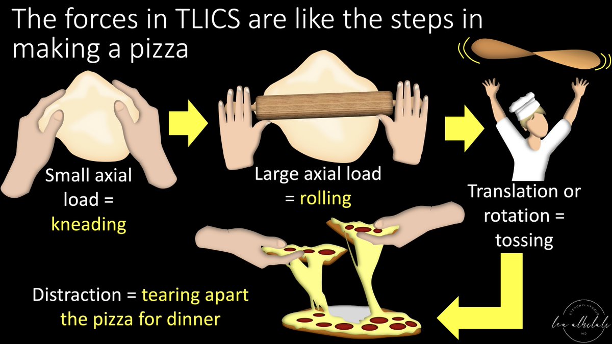
2/TLICS scores a fx on (1) morphology & (2) posterior ligamentous complex injury. Let's start w/morphology. TLICS scores severity like the steps to make & eat a pizza:

Mild compression (kneading), strong compression (rolling), rotation (tossing), & distraction (tearing in) Image
3/At the most mild, w/only mild axial loading, you get the simplest fx, a compression fx—like a simple long bone fx--worth 1 pt.

This is like when you just start to kneading the dough. There's pressure, but not as much as with a rolling pin! Image
Read 13 tweets

Did Thread Reader help you today?

Support us! We are indie developers!


This site is made by just two indie developers on a laptop doing marketing, support and development! Read more about the story.

Become a Premium Member ($3/month or $30/year) and get exclusive features!

Become Premium

Don't want to be a Premium member but still want to support us?

Make a small donation by buying us coffee ($5) or help with server cost ($10)

Donate via Paypal

Or Donate anonymously using crypto!

Ethereum

0xfe58350B80634f60Fa6Dc149a72b4DFbc17D341E copy

Bitcoin

3ATGMxNzCUFzxpMCHL5sWSt4DVtS8UqXpi copy

Thank you for your support!

Follow Us on Twitter!

:(