Liza Quintana, MD Profile picture
Mar 19, 2020 6 tweets 5 min read Read on X
Breast biopsy for calcs. What is your diagnosis? Would you do stains? Since I’m asking that means I did 😉🔬 #breastpath @BIDMCpath @CollinsLauraC @jennpincus @RanaAldreesMD @tlabiano @lara_pijuan @ADamronMD @ariella8 @kis_lorand @TheISBP ImageImageImageImage
Thanks for all the comments! Here are the stain. All (including p120, not pictured) were consistent with a lobular phenotype. ImageImage
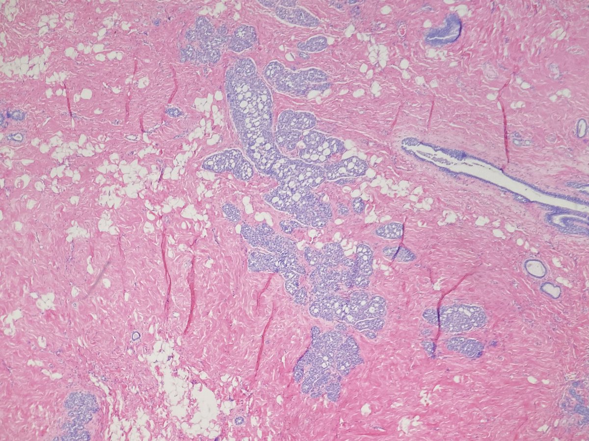
There were some hints in the background with areas of classical LCIS. ImageImage
This is a nice example of florid LCIS with comedo necrosis and associated calcifications. The nuclei aren't pleomorphic enough for me to call PLCIS; I would classify these are type B cells. Exc was recommended and showed more florid and classical LCIS with no invasive component.
We have limited info on the natural history of PLCIS and FLCIS. Per the who 25-60% of cases on core are upgraded to carcinoma on exc therefore exc is recommended.
Here is an example of PLCIS with associated invasive lobular I had a couple years ago.

• • •

Missing some Tweet in this thread? You can try to force a refresh
 

Keep Current with Liza Quintana, MD

Liza Quintana, MD Profile picture

Stay in touch and get notified when new unrolls are available from this author!

Read all threads

This Thread may be Removed Anytime!

PDF

Twitter may remove this content at anytime! Save it as PDF for later use!

Try unrolling a thread yourself!

how to unroll video
  1. Follow @ThreadReaderApp to mention us!

  2. From a Twitter thread mention us with a keyword "unroll"
@threadreaderapp unroll

Practice here first or read more on our help page!

More from @LizaMQuintana

Nov 20, 2020
1/ #BreastRadPath with @DrJordanaP

For this tweetorial we are going to talk about how we evaluate the biomarkers ER, PR, and HER2.

@BIDMCpath @BIDMC_BreastImg @CollinsLauraC @ADamronMD @GuzmanArocho @XeniaParisi @MarcosLepeMD @DrGeeONE @smlungpathguy
2/ The biomarkers provide predictive information (how a patient may respond to targeted therapy) as well as prognostic information. It helps to organize patients into treatment groups that follow different algorithms and guidelines.
3/ Biomarkers are evaluated in routine clinical mgmt of patients with breast cancer. The 3 routinely tested:

Hormone receptors (HR): Estrogen receptor (ER) and Progesterone receptor (PR)
&
Human epidermal growth factor receptor 2 (HER2)

(chart from: bit.ly/3lPbqoY)
Read 18 tweets
Oct 14, 2020
#breastradpath with @DrJordanaP

For #BreastCancerAwarenessMonth month we want to review the process from imaging to diagnosis of breast cancer. 1/ Image
It starts when a patient comes in for a screening or diagnostic mammogram. Here is @DrJordanaP reviewing #mammograms in the reading room. 2/ Image
Please review screening modalities in this amazing tweetorial from last week from @DrJordanaP! 3/

Read 24 tweets
Sep 28, 2020
Case 3 #breastradpath with @DrJordanaP

44 yo woman. On req: "faint grouped microcalcs." #breastimagers separate calcs and no calcs cores into separate containers (so helpful!). There was only one block of cores with calcs. Here are the #breastpath images. Thoughts? Next steps? ImageImage
You are all thinking the way I did! For cases with calcs, I always review the imaging, and in particular the specimen radiograph, to see the morphology of the calcs I should look for. Check out the imaging from @DrJordanaP 👇

The tiny calc in the initial levels of the CNB are not the same as the calcs seen on imaging. We need to find those calcs --> LEVELS! (I haven't heard it called steps before! I like!) #breastradpath correlation is so important here! ImageImage
Read 11 tweets
Sep 17, 2020
Hi #pathology tweeples! I just realized that the callus I have on my pinky is from using it to steady my hand as I dot 😂(with my green pen of course 😉). I must dot A LOT! 💪🔬

I have some #path (mostly) dotting related questions out of curiosity.

@BIDMCpath Image
The #pathology tweeples have very strong opinions about dotting pen color! My tweet is inspired by this from @Chucktowndoc
Does anyone else have a dotting pen-induced callus? (Just me? I hope I'm not alone. 🤞😊)
Read 11 tweets
Sep 15, 2020
#breastradpath with @DrJordanaP

We are excited to share the first case in our #breastimaging and #breastpath correlation series! This case highlights challenges of imaging/management and the pathologic diagnosis.

@BIDMC_BreastImg @BIDMCpath #radres #radfellows Image
56 yo woman with left breast focal asymmetry and calcifications. Screening mammogram. @DrJordanaP Image
Diagnostic mammogram and ultrasound were performed. A presumed ultrasound correlate was found with calcs and vascularity. What is the next step? What BI-RADS would give? @DrJordanaP ImageImage
Read 17 tweets
Mar 10, 2020
Breast excision. What is your diagnosis? Would you do stains? #breastpath ImageImageImage
Thanks everyone! This is LCIS involving collagenous spherulosis. I did IHC. The e-cad was stronger than I expected but showed granular staining so I followed up with p120 and beta-catenin to illustrate the different stains.
ImageImageImageImage
Read 5 tweets

Did Thread Reader help you today?

Support us! We are indie developers!


This site is made by just two indie developers on a laptop doing marketing, support and development! Read more about the story.

Become a Premium Member ($3/month or $30/year) and get exclusive features!

Become Premium

Don't want to be a Premium member but still want to support us?

Make a small donation by buying us coffee ($5) or help with server cost ($10)

Donate via Paypal

Or Donate anonymously using crypto!

Ethereum

0xfe58350B80634f60Fa6Dc149a72b4DFbc17D341E copy

Bitcoin

3ATGMxNzCUFzxpMCHL5sWSt4DVtS8UqXpi copy

Thank you for your support!

Follow Us!

:(