Pathologist #breastpath & #cytopath | #breastradpath| Director, Breast Path Fellowship | @BIDMCpath @BIDMChealth @harvardmed | RTs ≠ Endorsements
2 subscribers
How to get URL link on X (Twitter) App


 2/ The biomarkers provide predictive information (how a patient may respond to targeted therapy) as well as prognostic information. It helps to organize patients into treatment groups that follow different algorithms and guidelines.

 It starts when a patient comes in for a screening or diagnostic mammogram. Here is @DrJordanaP reviewing #mammograms in the reading room. 2/


 You are all thinking the way I did! For cases with calcs, I always review the imaging, and in particular the specimen radiograph, to see the morphology of the calcs I should look for. Check out the imaging from @DrJordanaP 👇
https://twitter.com/DrJordanaP/status/1311274647619428352?s=20

 The #pathology tweeples have very strong opinions about dotting pen color! My tweet is inspired by this from @Chucktowndoc
 https://twitter.com/Chucktowndoc/status/1299485866566529024?s=20

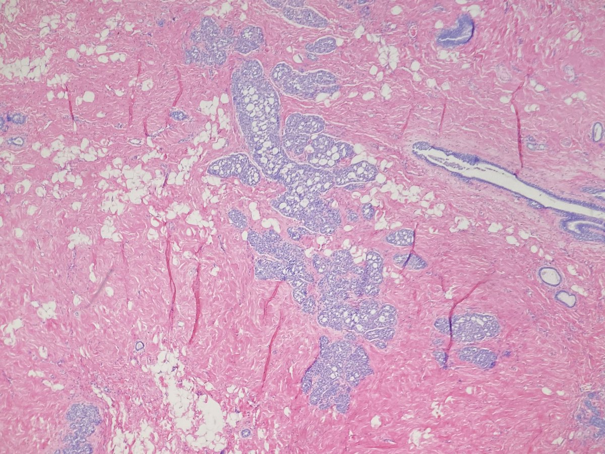
 56 yo woman with left breast focal asymmetry and calcifications. Screening mammogram. @DrJordanaP


 Thanks for all the comments! Here are the stain. All (including p120, not pictured) were consistent with a lobular phenotype.


 Thanks everyone! This is LCIS involving collagenous spherulosis. I did IHC. The e-cad was stronger than I expected but showed granular staining so I followed up with p120 and beta-catenin to illustrate the different stains.