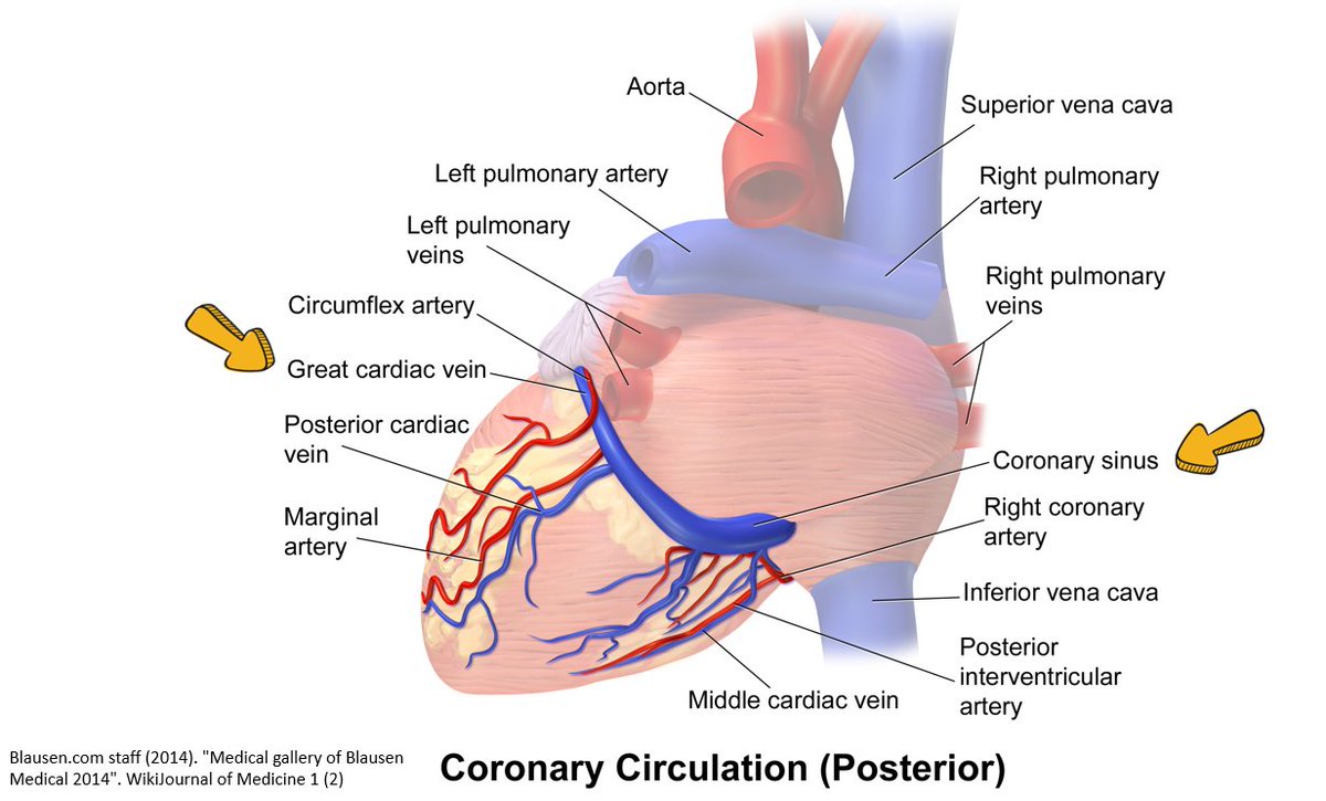
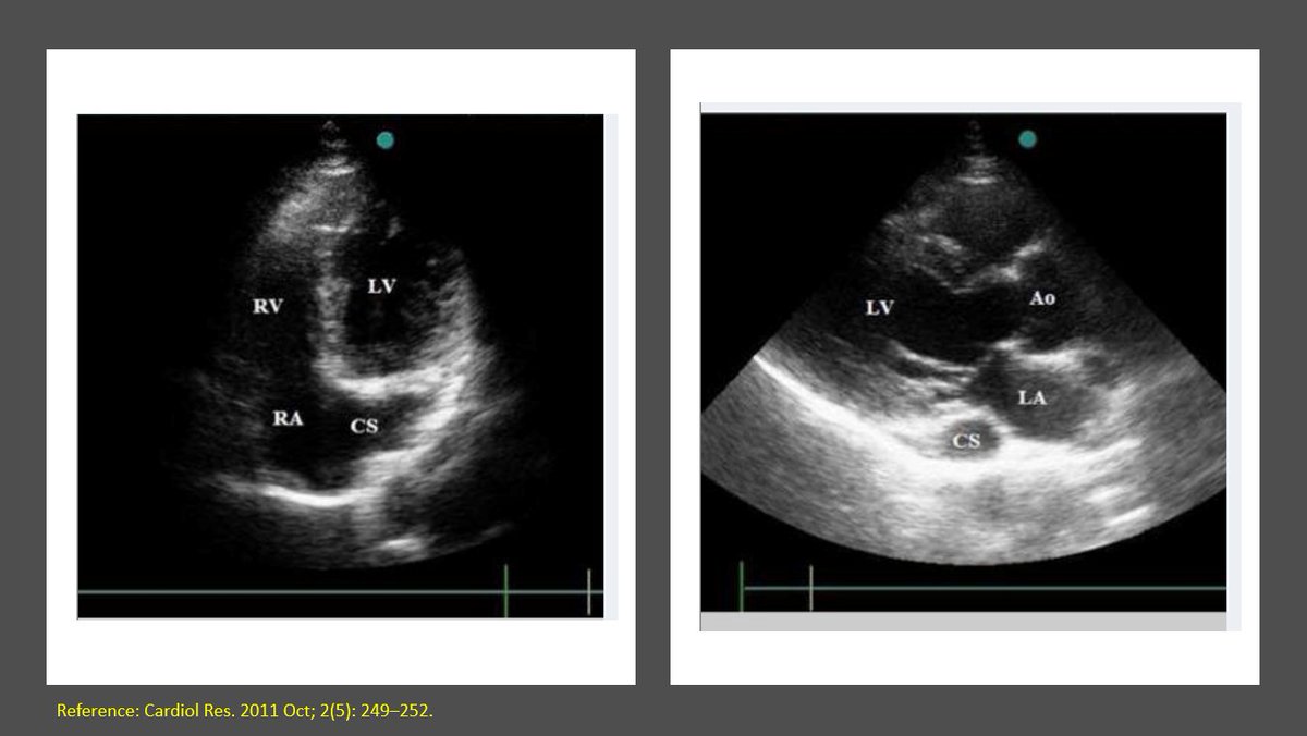
Dr. X is rounding on an ESRD pt who initially presented with dyspnea after missing a dialysis (HD) session; underwent dialysis in the hospital. Pt asymptomatic at the time of exam and lung #ultrasound revealed 👇 Further story in thread #MedEd
Info on various lung scan zones👇
Why can't we get more fluid out of a hypervolemic patient? Dr. X is perplexed and decides to more #POCUS Here is the IVC
#VExUS is not needed if IVC is small but just to make sure, hepatic and portal vein Doppler obtained - look normal!
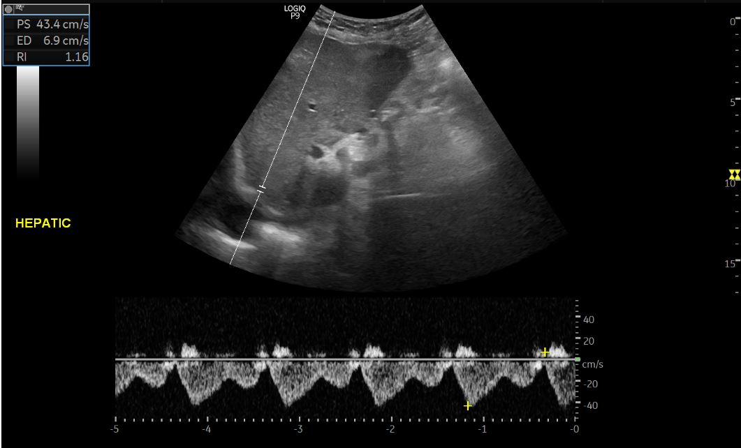
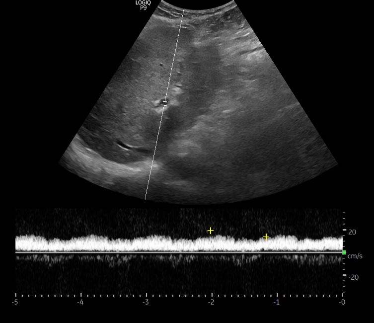
This time Dr. X does 8-zone #POCUS scan and finds similar images.
Pump function? diastolic dysfunction? bad mitral regurgitation?
Cardiac #POCUS performed. Not great windows but here is what Dr. X managed to get. Subcostal👇
Little bit of maneuvering shows some aortic valve calcification. Aortic stenosis? gradient not measured but seems to be opening, LV doesn't look obviously big/thick. Pt is 60+, kidney disease.
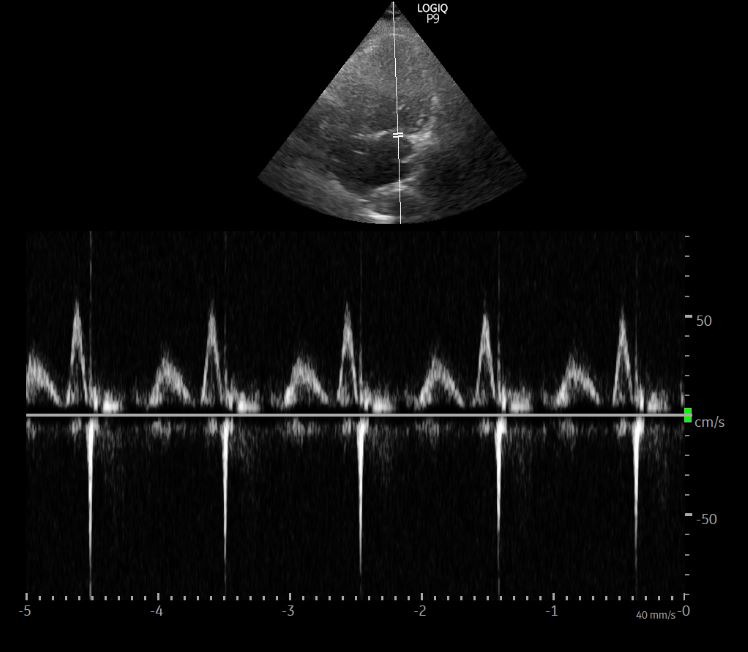
Impaired relaxation (A>E) but not unexpected in older people. Shouldn't fill the lung with B-lines. Tissue Doppler not performed.
Need a refresher on diastology? Refer to @Pocus101 's guide pocus101.com/how-to-measure…
Left lung images👇
Irregular pleural line suggestive of underlying lung disease (COVID negative, no symptoms). A CT chest was ordered.
Verdict: B-lines on lung #POCUS were likely not due to water. Pt referred to Pulmonologist.
In these cases, pleural line is irregular, show subpleural consolidations, may be thickened and some areas are spared.
Diffuse involvement as in this Pt can create confusion.
Any input @kyliebaker888 @siddharth_dugar @msiuba 🧐


