#breastradpath with @DrJordanaP
We are excited to share the first case in our #breastimaging and #breastpath correlation series! This case highlights challenges of imaging/management and the pathologic diagnosis.
@BIDMC_BreastImg @BIDMCpath #radres #radfellows
We are excited to share the first case in our #breastimaging and #breastpath correlation series! This case highlights challenges of imaging/management and the pathologic diagnosis.
@BIDMC_BreastImg @BIDMCpath #radres #radfellows

56 yo woman with left breast focal asymmetry and calcifications. Screening mammogram. @DrJordanaP 

Diagnostic mammogram and ultrasound were performed. A presumed ultrasound correlate was found with calcs and vascularity. What is the next step? What BI-RADS would give? @DrJordanaP 


Ultrasound-guided core needle biopsy was performed. Specimen radiograph done -- no calcs seen. Post-biopsy mammo shows clip near target. @DrJordanaP 


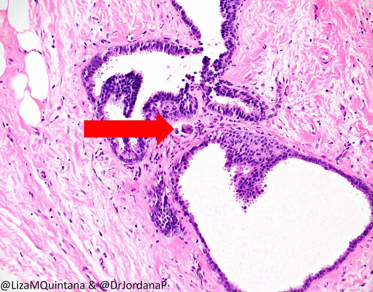
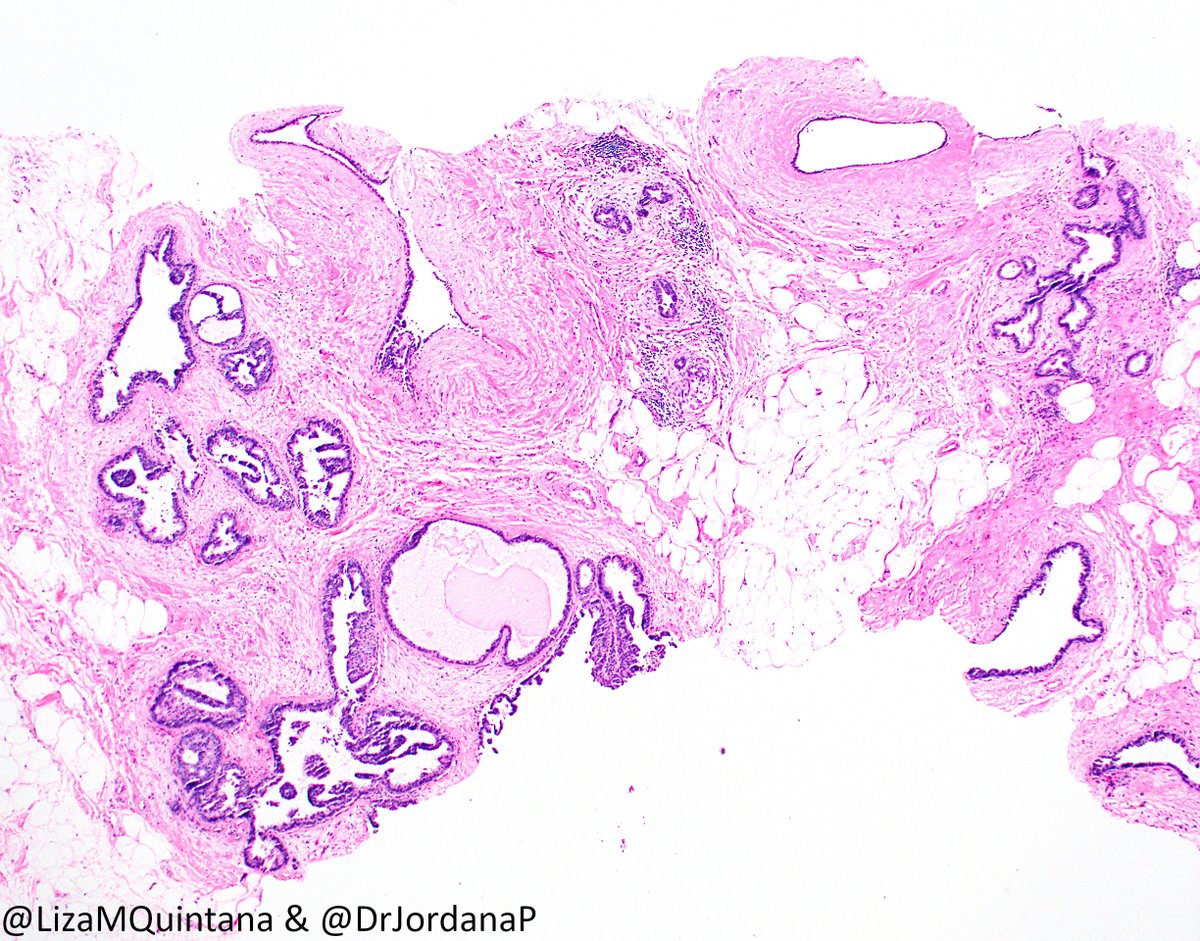
Great discussion everyone! This is a low-grade atypical proliferation (monotonous small nuclei). Based on how focal (quantity) and that it does not completely involve the spaces (quality) this is best cat as ADH (micropap and some rigid bridges) and FEA. Some CCC in background.
The distortion and periductal fibrosis raised the possibility of an associated sclerosing lesion (added in a note). Rare tiny calcs were seen (below) but were not detected in specimen radiograph, so additional levels wouldn't be helpful for calcs. 

So... is this concordant? What should next steps be? #breastimaging
https://twitter.com/DrJordanaP/status/1306004894407692289?s=20
From @DrJordanaP : Concerned about the initial finding - new focal asymm with calcs. ADH felt discordant, esp bc not sure how great our sample was (no calcs in specimen mg). So we did stereo and targeted calcs. Post-mg shows new bar clip near coil clip.
https://twitter.com/DrJordanaP/status/1306625726716624896?s=20
Path from biopsy #2 continued. Thoughts? Do we have the target? Summary teaching/take-home points to follow! 


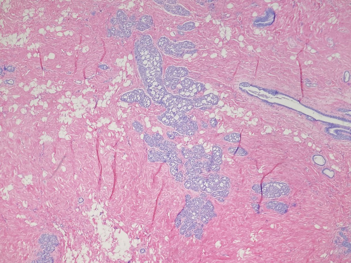
Thank you all for following along! Biopsy #2 showed spectrum of atypia -- FEA, through ADH, to areas with sufficient cyto and arch atypia for low-grade DCIS. There was periductal and stromal fibrosis associated with the atypia (same on excision) -- the asymmetry seen on imaging.
#breastradpath correlation for biopsy #2: concordant.
Here are our take home points for case #1!
@DrJordanaP


Here are our take home points for case #1!
@DrJordanaP


Some articles linked 👇
https://twitter.com/DrJordanaP/status/1307460743952990215
Why do a second biopsy? See @DrJordanaP response to the great question from @ADamronMD
👇
👇
https://twitter.com/DrJordanaP/status/1307475792700596224?s=20
• • •
Missing some Tweet in this thread? You can try to
force a refresh


