Case 3 #breastradpath with @DrJordanaP
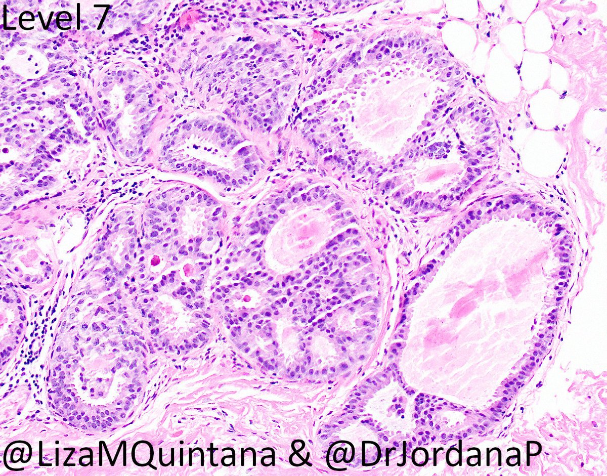
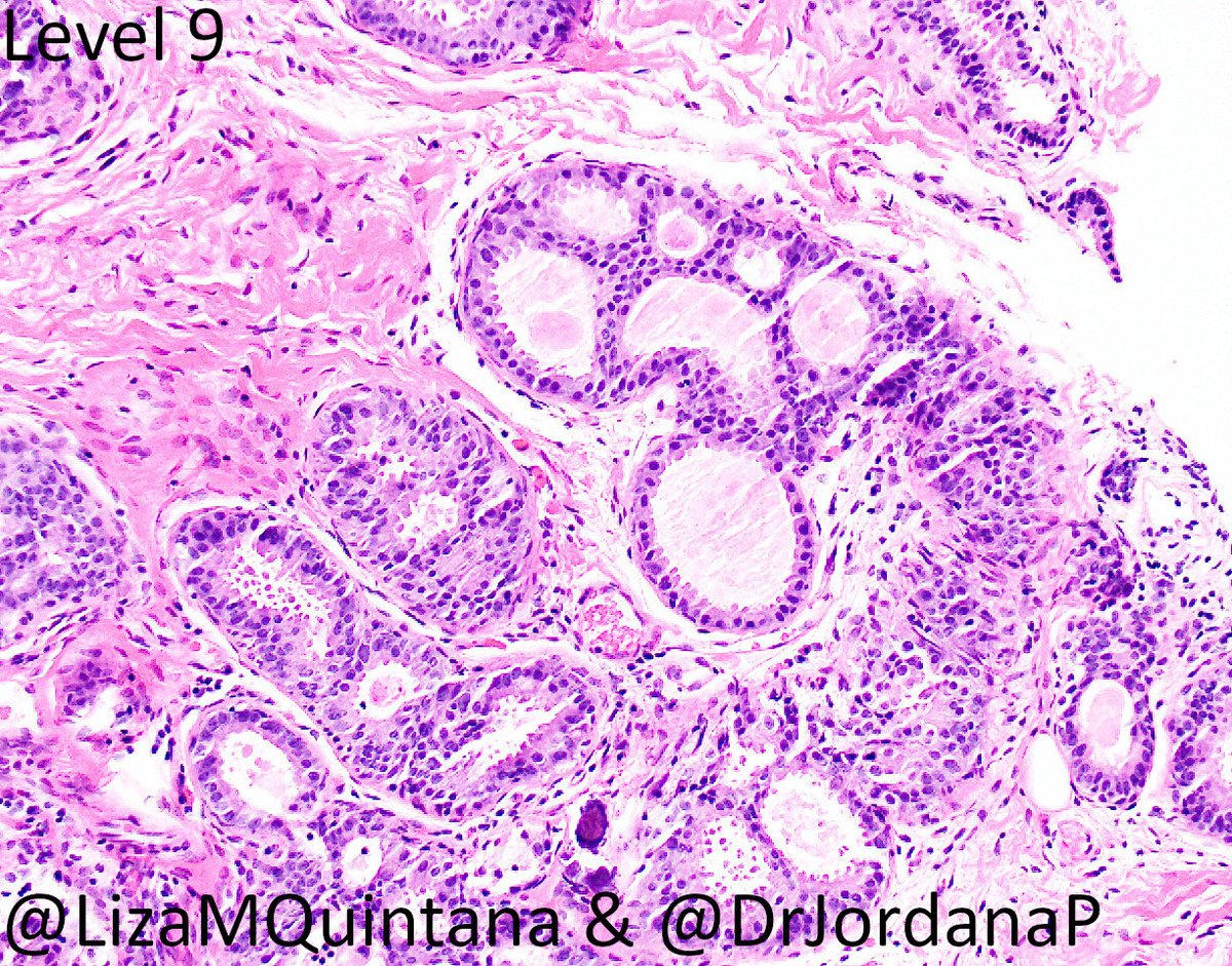
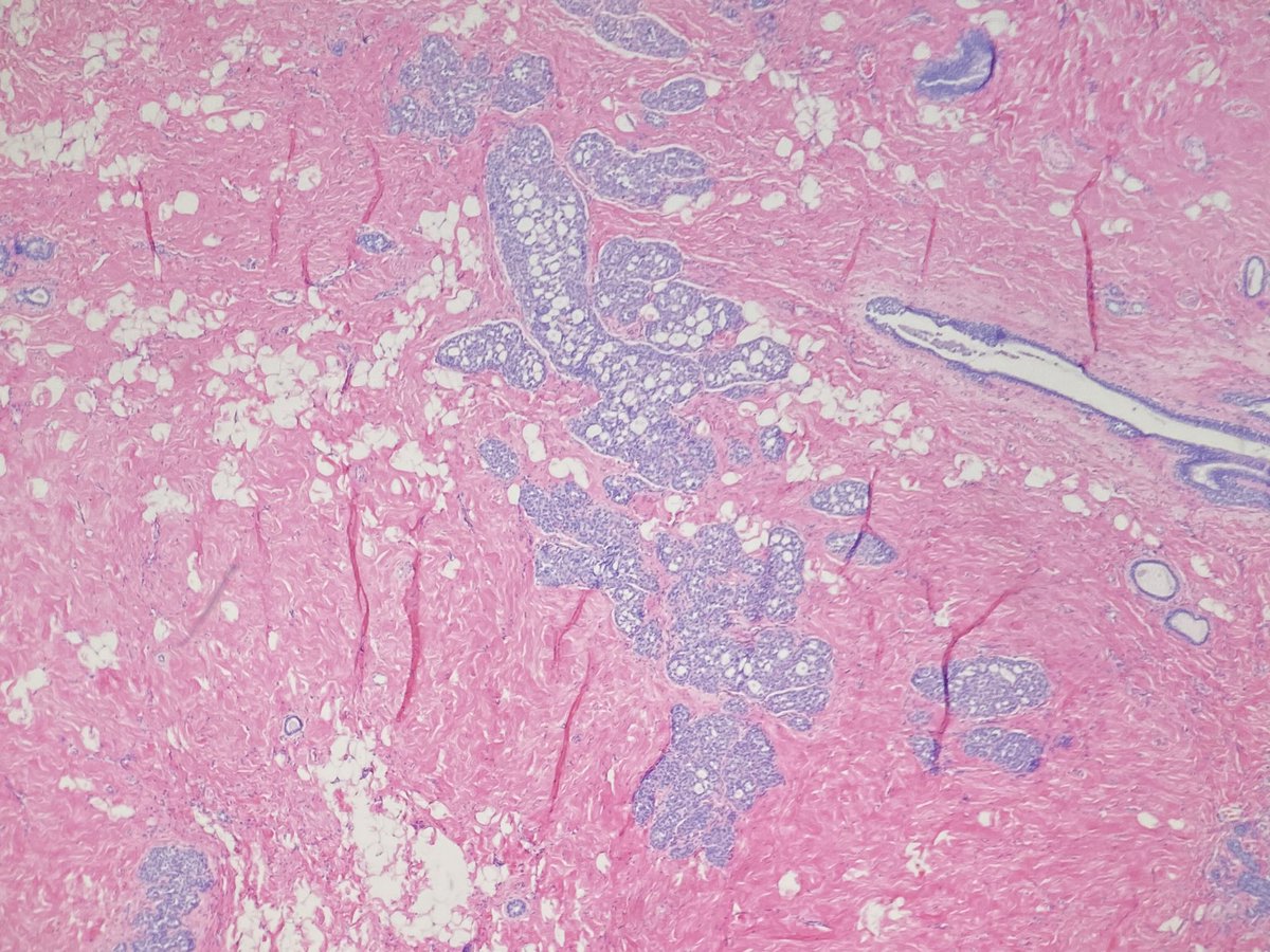
44 yo woman. On req: "faint grouped microcalcs." #breastimagers separate calcs and no calcs cores into separate containers (so helpful!). There was only one block of cores with calcs. Here are the #breastpath images. Thoughts? Next steps?

44 yo woman. On req: "faint grouped microcalcs." #breastimagers separate calcs and no calcs cores into separate containers (so helpful!). There was only one block of cores with calcs. Here are the #breastpath images. Thoughts? Next steps?


You are all thinking the way I did! For cases with calcs, I always review the imaging, and in particular the specimen radiograph, to see the morphology of the calcs I should look for. Check out the imaging from @DrJordanaP 👇
https://twitter.com/DrJordanaP/status/1311274647619428352?s=20
The tiny calc in the initial levels of the CNB are not the same as the calcs seen on imaging. We need to find those calcs --> LEVELS! (I haven't heard it called steps before! I like!) #breastradpath correlation is so important here! 


I'm curious, for the pathologists, do you call it "steps" or "levels"?
Path discussion: The span of atypia is 4.5 mm. For me, the quality of the prolif falls short of LG DCIS. In these cases we diagnose as: severely atypical intraductal proliferation bordering on LG DCIS arising in association with FEA; calcs are associated with the atypia.
This highlights how important it is for pathologists to review imaging/spec radiograph esp for calcs! We need to ensure that our levels are rep of imaging. In this case initial levels only showed a bit of focal ?FEA with a small calc. You all knew to get levels/steps 😊🔬
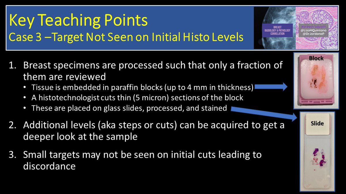
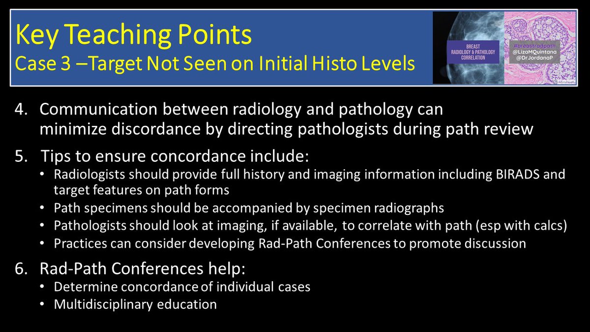
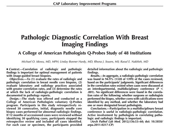
Here are some teaching points about this case and a few papers highlighting importance of #breastradpath concordance and utility of a multidisciplinary conference (including one by our very own @BIDMC_BreastImg group). 



@threadreaderapp unroll
• • •
Missing some Tweet in this thread? You can try to
force a refresh


