Hello subfield-fans! Last week's #SubfieldWednesday topic was the layered composition of the hippocampal subfields. We learned that the subfields contain three major cellular layers which makes them a part of the allocortex.
#SubfieldWednesday (1/n)
#SubfieldWednesday (1/n)

@thomcat992 replied that the hippocampus is archicortex, which is also correct! Archicortex is a type of allocortex.
#SubfieldWednesday (2/n)
#SubfieldWednesday (2/n)
Now what about the entorhinal cortex (ERC)? The ERC has six layers, so does that make it neocortex (also known as the isocortex)?
#SubfieldWednesday (3/n)
#SubfieldWednesday (3/n)
@DrNeuroChic correctly replied that the ERC is mesocortex, which refers to a transitional cortex that does not have the same layer features as neocortex!
#SubfieldWednesday (4/n)
#SubfieldWednesday (4/n)
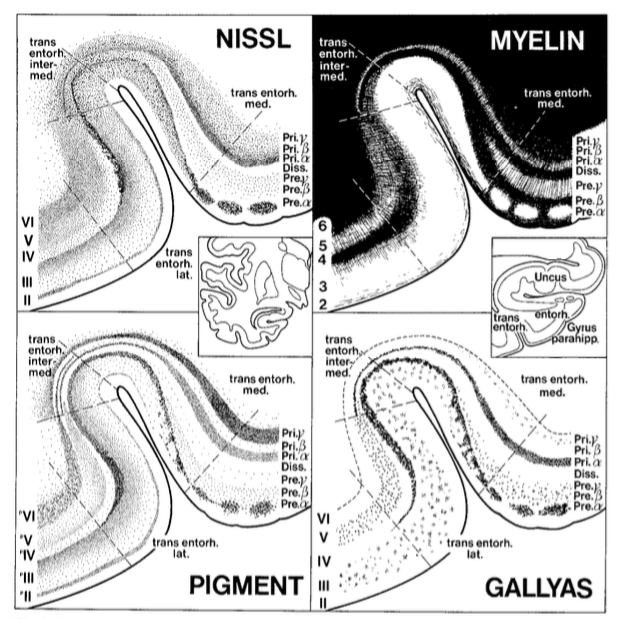
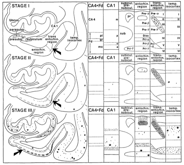
Insausti describes ERC (as well as the pre- and parasubiculum) as being a part of a specific transitional cortex called periallocortex.
#SubfieldWednesday (5/n)
#SubfieldWednesday (5/n)
A common feature of all components of the periallocortex is the presence of a cell free zone, parallel to the pial surface, which receives the name of lamina dissecans.
#SubfieldWednesday (6/n)
#SubfieldWednesday (6/n)

Insausti further describes perirhinal (PRC) and parahippocamal cortex (PHC) as being a part of a different transitional cortex called proisocortex.
According to Insausti, the proisocortex does not fulfill all the layering features of neocortex.
#SubfieldWednesday (7/n)
According to Insausti, the proisocortex does not fulfill all the layering features of neocortex.
#SubfieldWednesday (7/n)
"The proisocortex shows six layers, ...., but retain some of the periallocortical features such as prominent layers II and V, the lack or a thin layer IV, and an overall lesser columnarity than the isocortex." (Insausti et al., Front Neuroanatomy, 2017)
#SubfieldWednesday (8/n)
#SubfieldWednesday (8/n)
@madeopyj ventured a guess that medial and lateral ERC might be composed of different kinds of cortex. Indeed, Insausti describes medial-lateral gradients within the ERC, such that lateral ERC is more similar to the proisocortex of the PRC.
#SubfieldWednesday (9/n)
#SubfieldWednesday (9/n)
That's all for this week! Thanks to all who participated in the "quiz"! See you next week!
#SubfieldWednesday (end)
#SubfieldWednesday (end)
I should clarify that *some* of the subfields have three layers:
The dentate gyrus, cornu ammonis, and subiculum proper
See thread below regarding which subfields have more than three layers!
The dentate gyrus, cornu ammonis, and subiculum proper
See thread below regarding which subfields have more than three layers!
@threadreaderapp unroll
• • •
Missing some Tweet in this thread? You can try to
force a refresh


