Happy #SubfieldWednesday! After a two-week hiatus we are returning to our quiz about the mysterious transentorhinal cortex!

This quiz even stumped some of us at @hipposubfields headquarters! We had to contact a neuroanatomist to confirm which answer is correct! (or at least "the most correct")
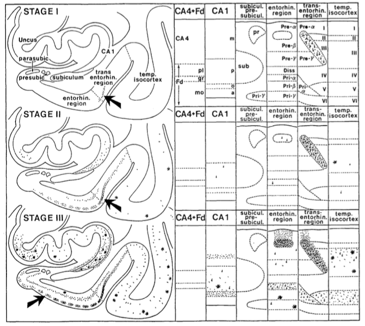
Braak and Braak (1985) originally described the transentorhinal cortex as a 'transition region between entorhinal cortex and temporal isocortex. This rules out answer A (part of ERC) Figure 3. from Braak H, Bra...
So, if it's not part of ERC, is it part of PRC?
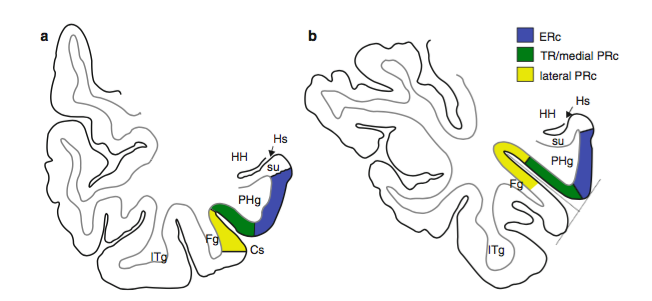
Kivissari et al., (2013) describe transentorhinal (labeled TR below) as the medial part of PRC, and separate from ERC. Figure 19.4 from Kivisaari,...
According to Augustinack et al. (NeuroImage, 2013):

"[Perirhinal] area 35 and transentorhinal are somewhat synonymous terms."
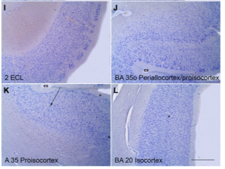
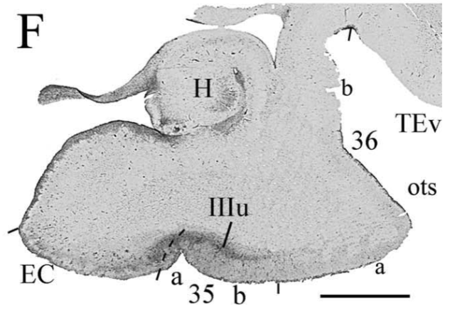
Insausti et al., (Front NeuroAnat, 2017) concur that the proisocortex of BA35 is the same as Braak and Braak's transentorhinal cortex (see panel K). Adapted from Figure 4 of In...
Finally, Ding & Van Hoesen (Human Brain Mapping, 2010) describes tau lesions in BA35, which is consistent with overlap between BA35 and transentorhinal cortex. Tau lesions are present in ...
So, the correct answer is C: "Roughly the same as BA35"
Also note, according to both Insausti and Ding, that the location of BA35/transentorhinal cortex can vary in relationship to the collateral sulcus, depending on the depth of that sulcus!
We would also like to mention that there might be some disagreement among neuroanatomists about the definition of this area.

In fact, in the next phase of our harmonization effort, we hope to reconcile these different groupings/definitions.

• • •

Missing some Tweet in this thread? You can try to force a refresh
 

Keep Current with Hippocampal Subfield Group

Hippocampal Subfield Group Profile picture

Stay in touch and get notified when new unrolls are available from this author!

Read all threads

This Thread may be Removed Anytime!

PDF

Twitter may remove this content at anytime! Save it as PDF for later use!

Try unrolling a thread yourself!

how to unroll video
  1. Follow @ThreadReaderApp to mention us!

  2. From a Twitter thread mention us with a keyword "unroll"
@threadreaderapp unroll

Practice here first or read more on our help page!

More from @hipposubfields

21 Oct
Last week we received over 100 (!) responses on our quiz about which hippocampal subfield accumulates tau pathology earliest in the disease.

#SubfieldWednesday (1/n)
This week we will explain that the correct answer for Alzheimer's disease (but not all types of dementia) is CA1!

#SubfieldWednesday (2/n)
Work from Braak & Braak (1991) showed that CA1 develops tau pathology before the other subfields.

#SubfieldWednesday (3/n) Cartoon drawing of the medi...
Read 9 tweets
8 Oct
Hello subfield-fans! Last week's #SubfieldWednesday topic was the layered composition of the hippocampal subfields. We learned that the subfields contain three major cellular layers which makes them a part of the allocortex.

#SubfieldWednesday (1/n) Table 1 from: Insausti, Ric...
@thomcat992 replied that the hippocampus is archicortex, which is also correct! Archicortex is a type of allocortex.

#SubfieldWednesday (2/n)
Now what about the entorhinal cortex (ERC)? The ERC has six layers, so does that make it neocortex (also known as the isocortex)?

#SubfieldWednesday (3/n)
Read 12 tweets
1 Oct
Better late than never! (It's still Wednesday in some timezones!) This week's #SubfieldWednesday will be a bit briefer than normal.

#SubfieldWednesday (1/n)
Some of you might have been surprised last week when we described the hippocampal subfields as layered structures.

#SubfieldWednesday (2/n)
Even though the hippocampus sometimes gets grouped together with subcortical structures like the thalamus and basal ganglia, the dentate gyrus, CA fields, and subiculum are all part of a special type of cortex called the allocortex!

#SubfieldWednesday (3/n)
Read 7 tweets
23 Sep
It's Wednesday...so you know what that means! It's time for #SubfieldWednesday! 🥳📢

(1/n)
Two weeks ago we posted about whether you can visualize subfields on in vivo MRI. Here is a link if you missed it!


#SubfieldWednesday (2/n)
You might recall that the hippocampus is a layered structure (just like the neocortex!) and that some of the layers have different contrast properties due to different amounts of myelination and cell densities

#SubfieldWednesday (3/n) post-mortem, ultra-high fie...Drawing of the different la...
Read 18 tweets
9 Sep
This question for this week's #SubfieldWednesday is:

Can you visualize subfields using in vivo magnetic resonance imaging (MRI)? (1/n)
Answer:

Yes, there are specific anatomical features in MRI that we can use to identify different hippocampal subfields (2/n)

#SubfieldWednesday
How is this possible? If subfields are defined based on cell types, cell size and density, layer thickness, etc.? Neuroanatomists typically define subfields based on special dyes that stain the cell bodies (e.g. Nissl stain as shown below) #SubfieldWednesday (3/n) ImageImage
Read 15 tweets
2 Sep
Drum roll please...it's time for our very first #SubfieldWednesday!
One thing you might be wondering is: How many subfields are there in the human hippocampus and what are they called??

The answer actually depends on who you ask! Let me explain more!

#SubfieldWednesday
Most people who study the hippocampal subfields with high resolution MRI recognize CA1, CA2, CA3, CA4, dentate gyrus (DG), and the subiculum as the major subfields of the hippocampus.

That means there are five...right?

#SubfieldWednesday Image
Read 10 tweets

Did Thread Reader help you today?

Support us! We are indie developers!


This site is made by just two indie developers on a laptop doing marketing, support and development! Read more about the story.

Become a Premium Member ($3/month or $30/year) and get exclusive features!

Become Premium

Too expensive? Make a small donation by buying us coffee ($5) or help with server cost ($10)

Donate via Paypal Become our Patreon

Thank you for your support!

Follow Us on Twitter!