The craziness of the past week has settled down and #Breastradpath with @LizaMQuintana is back with Case 4!

53 yo had a screening MG. Maybe some new calcs or distortion in rt breast (arrow, 3D not shown)? #radres #radfellows -which images would you want during diag work-up?? Image
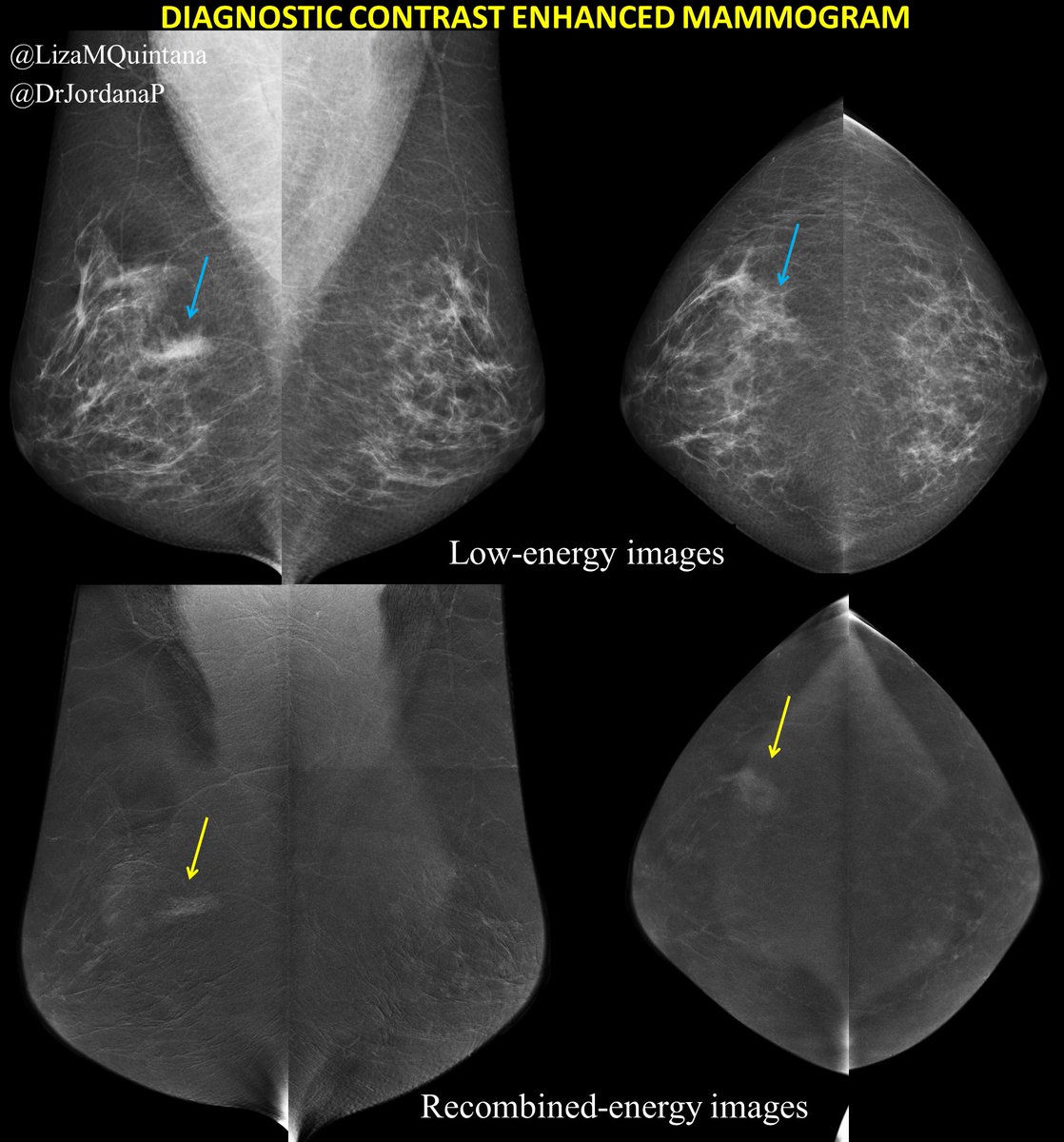
She returned for diag work-up. Common diag w/u is mag views for calcs and tomo spots/90 for poss AD. But we decided to start with #contrastmammo & use enhancement to triage. What do you think? Based on these images alone, does it change your level of concern???? Ddx??
Rather than showing you other diag images, which weren't that helpful, I'm showing the US. Could this be a correlate??? Does it change your level of concern?? Ddx??
Share thoughts below.... more to come tomorrow! Image
We thought the US was a fine correlate & decided to recommend US-guided bx. We coded the case BI-RADS 4B bc our Ddx included PASH or ILC (was super similar to Case 2 in our series). The clip correlated perfectly with the enhancement. Check out Case 2 👇

Representative path slides are included. Hey there #breastpath what do you think??? Share your thoughts below! ImageImageImage
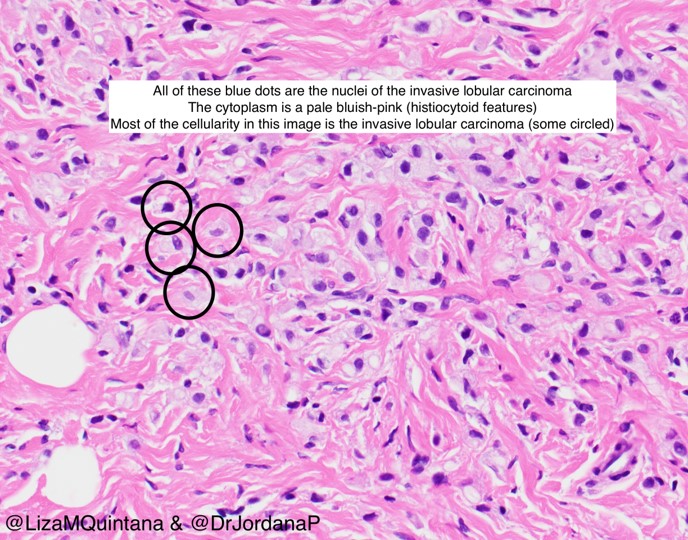
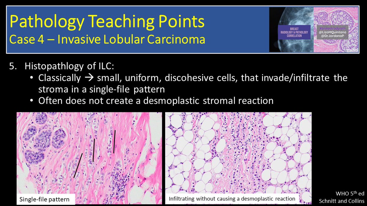
Nice job everyone! Turns out this case was NOT pash (like Case 2) but actually was ILC with histiocytoid features. You all got it right!
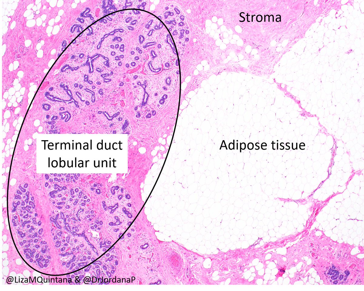
Normal stroma is on the left and the ILC is on the right. Notice how many more nuclei there are in the ILC (blue dots) indicating way more cells. They are also extending outside the lobule. The # of cells and growth pattern are how we know it's invasive lobular ImageImage
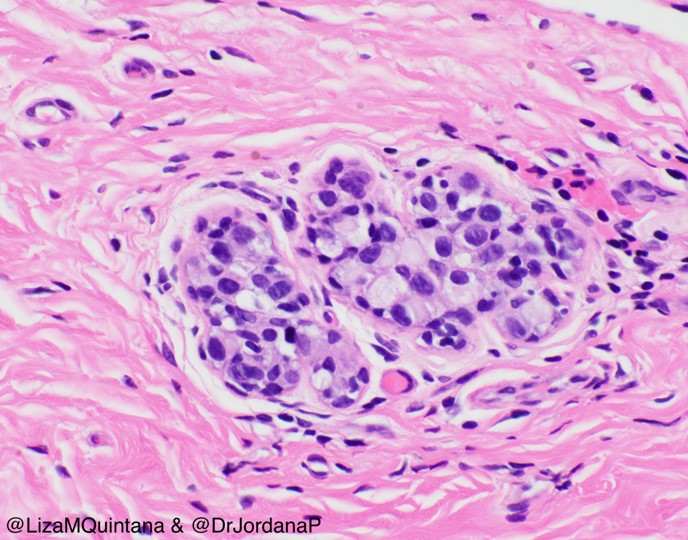
Here is an example of lobular neoplasia-many more cells but they are confined to the acinus of the lobule. Atypical lobular hyperplasia (ALH) & Lobular carcinoma (LCIS) are differentiated based on # of cells in acinus. More cells = LCIS, a non-obligate precursor to ILC. Image
For a refresher of normal anatomy - check out the tweetorial on male breast disease

Image
Teaching points and relevant articles to come in the next few days...
This week's topic was invasive lobular cancer. Check out teaching points and articles below! ImageImageImage
Here are the radiology teaching points. #radres #radfellows - did you know that ILC and PASH are both part of the differential for developing asymmetry??

Great Radiographics article on ILC:
pubs.rsna.org/doi/10.1148/rg…

#BrImagingPubClub ImageImageImage

• • •

Missing some Tweet in this thread? You can try to force a refresh
 

Keep Current with Jordana Phillips

Jordana Phillips Profile picture

Stay in touch and get notified when new unrolls are available from this author!

Read all threads

This Thread may be Removed Anytime!

PDF

Twitter may remove this content at anytime! Save it as PDF for later use!

Try unrolling a thread yourself!

how to unroll video
  1. Follow @ThreadReaderApp to mention us!

  2. From a Twitter thread mention us with a keyword "unroll"
@threadreaderapp unroll

Practice here first or read more on our help page!

More from @DrJordanaP

21 Oct 20
1/For this tweetorial in our #breastradpath series w @LizaMQuintana we're going to focus on #malebreastcancer for male breast cancer awareness week. An important topic that doesn't get enough attention! #BCAM @MBCC_MHBT @BreastCancerMen @BreastCancerNow @BreastImaging @Breast360
2/But before we can talk about the male breast, we first need to review the female breast. The female breast is made of skin, breast fat, ducts, and lobules. The breast sits on the pectoralis major muscle of the chest wall
3/The female breast is like a tree in springtime. A group of buds on a tree are like breast lobules (one bud is called an acinus). The tree branches are like breast ducts. Can you see the similarities between the sakura cherry tree and the specimen of breast tissue below???
Read 18 tweets
5 Oct 20
1/ Let the tweetorials begin for our #breastradpath series with @LizaMQuintana! We're starting with the basics of screening mammography for #BCAM

Each screening mammo has 4 images-2 of the right breast and 2 of the left. If any abnormality is seen then we get more images
2/ There are 2 main areas on all mammograms - white areas are glandular tissue (part of breast that makes milk, yellow on image) & black areas are breast fat (blue). All women have both but in different proportions #breastdensity
3/ Check out what this looks like on the pathology slide. And just to give some definitions: adipose tissue=breast fat, terminal duct lobular unit=glandular tissue, stroma=supporting structure of breast #Breastpath
Read 18 tweets
21 Sep 20
Case2 of #breastradpath with @LizaMQuintana

40 yo with focal asymmetry in rt breast on screening mg (blue arrows). See descriptions below all images.

#radres #radfellows #breastimaging #breastpath #pathology #breastmulti @BIDMC_BreastImg @BIDMCpath @DrTedJames Screening mammogram from to...
Patient returned for diagnostic #contrastmammo which confirmed focal asymmetry (blue arrows) & showed non-mass enhancement (NME) on recombined images (yellow arrows). US showed correlate. USG core done and post-MG confirmed target (yellow arrows). Contrast enhanced mammogram...Targeted ultrasound of the ...US guided core biopsy was d...
Calling all breast docs, what is your differential diagnosis? BI-RADS? Next steps??
Read 9 tweets

Did Thread Reader help you today?

Support us! We are indie developers!


This site is made by just two indie developers on a laptop doing marketing, support and development! Read more about the story.

Become a Premium Member ($3/month or $30/year) and get exclusive features!

Become Premium

Too expensive? Make a small donation by buying us coffee ($5) or help with server cost ($10)

Donate via Paypal

Or Donate anonymously using crypto!

Ethereum

0xfe58350B80634f60Fa6Dc149a72b4DFbc17D341E copy

Bitcoin

3ATGMxNzCUFzxpMCHL5sWSt4DVtS8UqXpi copy

Thank you for your support!

Follow Us on Twitter!

:(