1/7
Here is a quick Tweetorial on Abdominal Aorta #POCUS for you all!
It’s a RULE IN study! Not a rule out ⚠️
Images from a forthcoming book chapter with @LukeFlower1 @icmteaching + @ICUltrasonica !
#FOAMed #FOAMcc #echofirst
#medtwitter
Hopefully we won’t see these?!
Here is a quick Tweetorial on Abdominal Aorta #POCUS for you all!
It’s a RULE IN study! Not a rule out ⚠️
Images from a forthcoming book chapter with @LukeFlower1 @icmteaching + @ICUltrasonica !
#FOAMed #FOAMcc #echofirst
#medtwitter
Hopefully we won’t see these?!

2/7
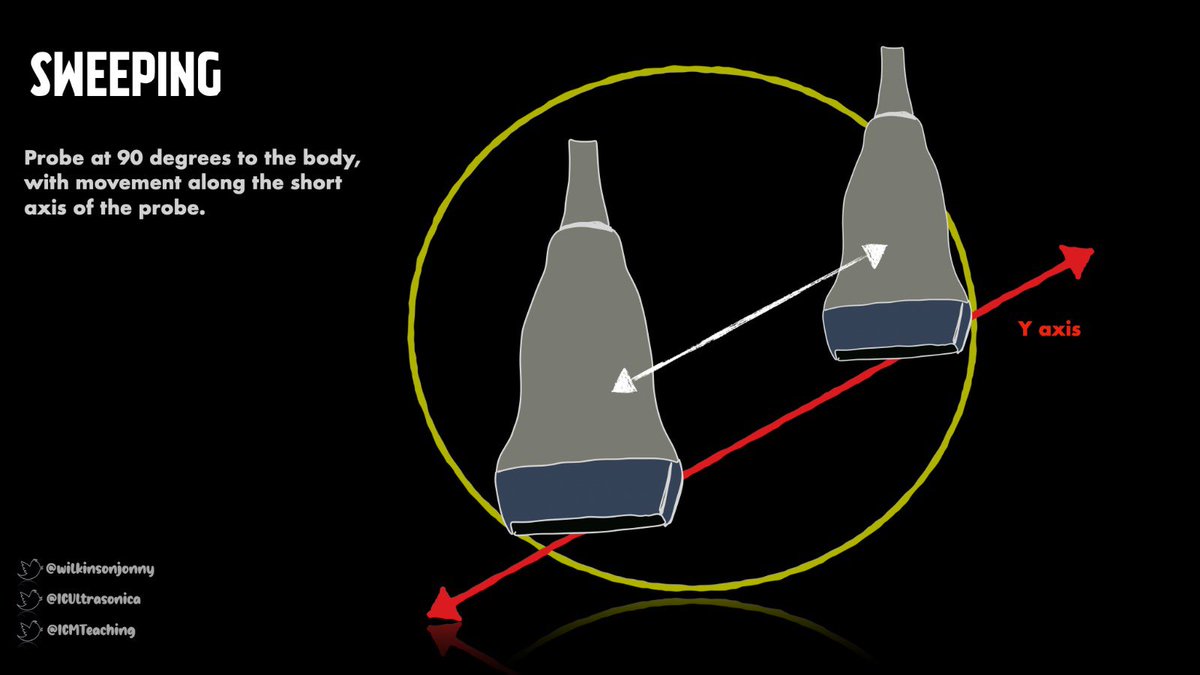
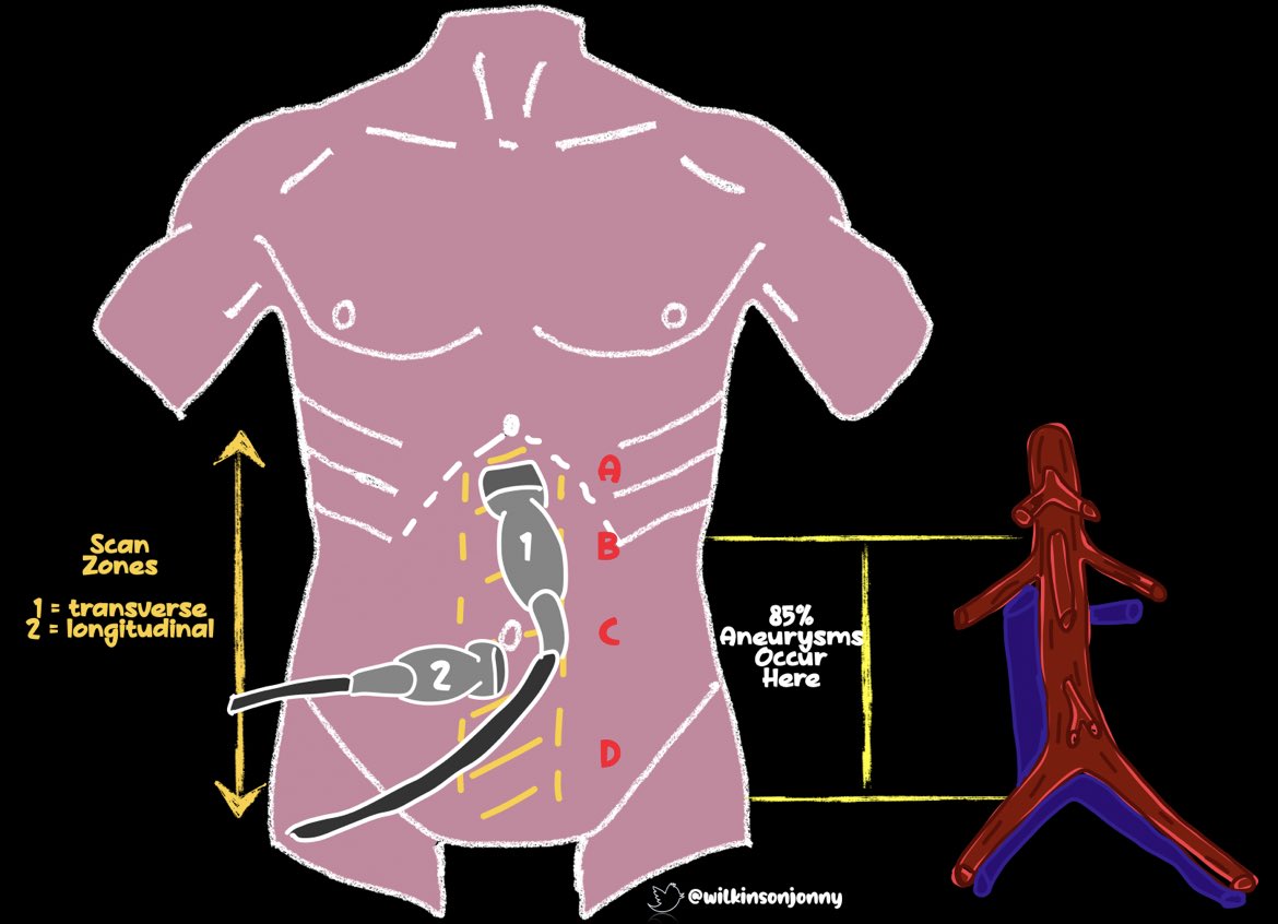
Apply careful firm pressure to displace pesky bowel gas. I start at the umbilicus; you can find the vertebral body easily here. You can then move up or down, tracing the vessel. The aim is to see as much of the vessel as you can. Marker - right (SAX) or to the head (LAX).
Apply careful firm pressure to displace pesky bowel gas. I start at the umbilicus; you can find the vertebral body easily here. You can then move up or down, tracing the vessel. The aim is to see as much of the vessel as you can. Marker - right (SAX) or to the head (LAX).
3/7
High Subxiphoid SAX
Find that vertebral body shadow again, you will see the aorta and IVC just above this. We are looking for the classic ‘seagull’ sign -
Hepatic artery and splenic artery = wings.
Coeliac trunk = body.
High Subxiphoid SAX
Find that vertebral body shadow again, you will see the aorta and IVC just above this. We are looking for the classic ‘seagull’ sign -
Hepatic artery and splenic artery = wings.
Coeliac trunk = body.

4/7
Upper Transverse View
Move caudally from the last view to get this one. Look for the vertebral body shadow. The aorta lies to the left and the IVC at 10 o’clock to this. The SMA is visible as a small pulsating shape at 11/12o’clock. Note the left renal vein hugs the aorta.
Upper Transverse View
Move caudally from the last view to get this one. Look for the vertebral body shadow. The aorta lies to the left and the IVC at 10 o’clock to this. The SMA is visible as a small pulsating shape at 11/12o’clock. Note the left renal vein hugs the aorta.

5/7
The Longitudinal View
2 major branches are apparent. The most cranial = coeliac trunk. Below this = SMA.
The renal arteries branch off below the SMA; not always visible. Right = posterior to the IVC. Other branches are subtle. Hence, CT best for I.D’ing prior to surgery.
The Longitudinal View
2 major branches are apparent. The most cranial = coeliac trunk. Below this = SMA.
The renal arteries branch off below the SMA; not always visible. Right = posterior to the IVC. Other branches are subtle. Hence, CT best for I.D’ing prior to surgery.

6/7
Bifurcation View (LAX / SAX)
Travelling further caudally, we get this view. We see the bifurcation into the common iliac arteries. IVC and spine lie posteriorly.
Diameter of the iliacs = 1.5cm in men; 1.2cm in women.
Bifurcation View (LAX / SAX)
Travelling further caudally, we get this view. We see the bifurcation into the common iliac arteries. IVC and spine lie posteriorly.
Diameter of the iliacs = 1.5cm in men; 1.2cm in women.


7/7
Measurement of the aorta at the umbilicus:
1.5-2cm - Normal
<3cm - discharge
3-4cm - annual scan
4-5cm - 3monthly scans
>5cm - refer to surgeon
>7cm - critical rupture risk!
Do LAX and SAX measurements - inner to inner wall, as per MASS study👍
pubmed.ncbi.nlm.nih.gov/12443589/
Measurement of the aorta at the umbilicus:
1.5-2cm - Normal
<3cm - discharge
3-4cm - annual scan
4-5cm - 3monthly scans
>5cm - refer to surgeon
>7cm - critical rupture risk!
Do LAX and SAX measurements - inner to inner wall, as per MASS study👍
pubmed.ncbi.nlm.nih.gov/12443589/
• • •
Missing some Tweet in this thread? You can try to
force a refresh