1-
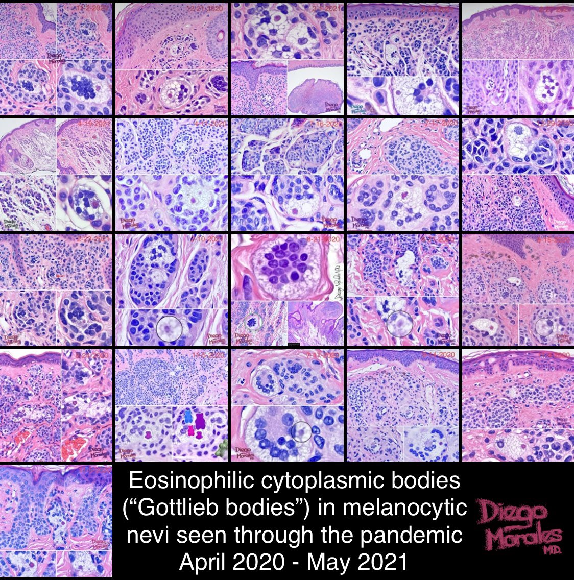
Eosinophilic cytoplasmic bodies in melanocytes.
Perhaps one of the most overlooked findings in #dermpath -according to me-. I will show all the cases I’ve found since ~April 2020 (around 20 cases) and say a few words about #Gottliebbodies 👇🏼
#dermtwitter #PathTwitter
Eosinophilic cytoplasmic bodies in melanocytes.
Perhaps one of the most overlooked findings in #dermpath -according to me-. I will show all the cases I’ve found since ~April 2020 (around 20 cases) and say a few words about #Gottliebbodies 👇🏼
#dermtwitter #PathTwitter

2-
🔴Gottlieb bodies
I don’t spend much more time looking for them, I only search when I see sebocyte-like melanocytes (not that rare)
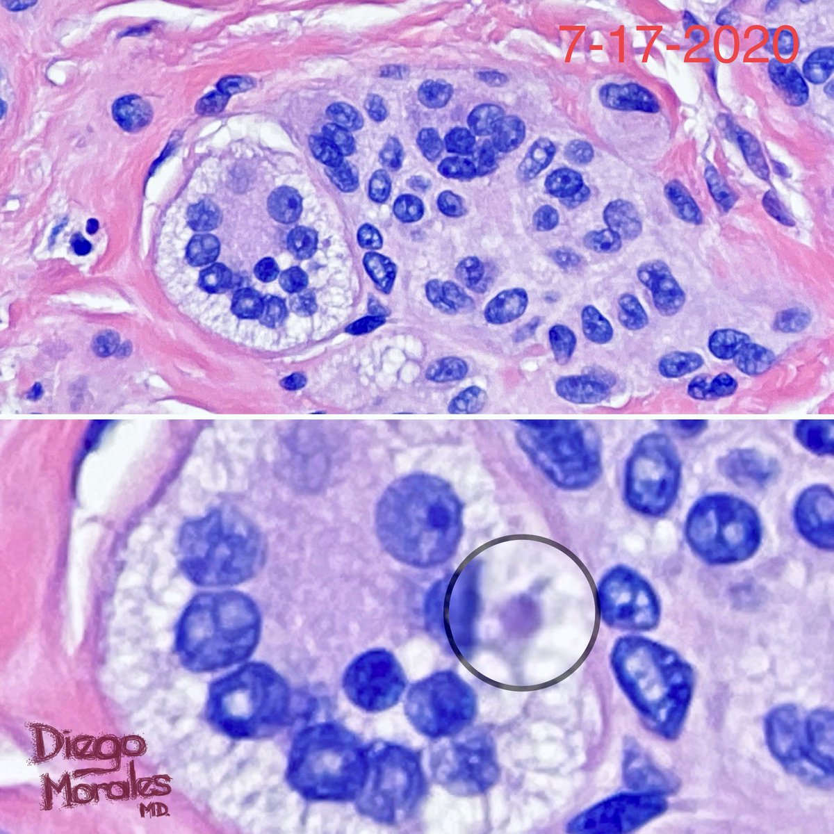
🏓 eosinophilic cytoplasmic body within an -usually-multinucleated vacuolated melanocyte
🏓 small incipient ones have basophilic appearance


🔴Gottlieb bodies
I don’t spend much more time looking for them, I only search when I see sebocyte-like melanocytes (not that rare)
🏓 eosinophilic cytoplasmic body within an -usually-multinucleated vacuolated melanocyte
🏓 small incipient ones have basophilic appearance


3-
🏓 larger fully developed ones have concentric lamination with eosinophilic staining
🏓 thought to represent degenerative changes in melanosomes, although this study doesn’t support such theory 👉🏼 pubmed.ncbi.nlm.nih.gov/21819442/


🏓 larger fully developed ones have concentric lamination with eosinophilic staining
🏓 thought to represent degenerative changes in melanosomes, although this study doesn’t support such theory 👉🏼 pubmed.ncbi.nlm.nih.gov/21819442/


4-
🏓 one or several bodies can be found within one melanocyte
🏓 seen in long-standing compound or intradermal melanocytic nevi
🏓 especially in nevi exhibiting congenital features


🏓 one or several bodies can be found within one melanocyte
🏓 seen in long-standing compound or intradermal melanocytic nevi
🏓 especially in nevi exhibiting congenital features


5-
🏓 not seen in melanomas, only when melanoma is associated with nevus
🏓 is a reliable #dermpathclue to the diagnosis of a nevus
🏓 I found one that looks like a #gummybear 😜
instagram.com/p/CHOCmgDjt7U/…


🏓 not seen in melanomas, only when melanoma is associated with nevus
🏓 is a reliable #dermpathclue to the diagnosis of a nevus
🏓 I found one that looks like a #gummybear 😜
instagram.com/p/CHOCmgDjt7U/…


6-
🏓 described by Geoffrey Gottlieb in 2002 @ Dermatopathol Practical & Conceptual; the only repository of such journal was Derm101.com, website deactivated by Galderma on 12/31/2019, which means its articles are hidden, unless you have the actual📙(I don’t 😪, you?)


🏓 described by Geoffrey Gottlieb in 2002 @ Dermatopathol Practical & Conceptual; the only repository of such journal was Derm101.com, website deactivated by Galderma on 12/31/2019, which means its articles are hidden, unless you have the actual📙(I don’t 😪, you?)


7-
🏓 by EM, they look like non-membrane bound structures, comprised of radiating filamentous elements with an electron-dense core, (only picture that is not taken by me)👉🏼 pubmed.ncbi.nlm.nih.gov/21819442/
@wshonpath
🏓 by EM, they look like non-membrane bound structures, comprised of radiating filamentous elements with an electron-dense core, (only picture that is not taken by me)👉🏼 pubmed.ncbi.nlm.nih.gov/21819442/
@wshonpath

8-
Thanks for making it to the end!
Look for them 🔴, no need to spend too much extra ⏳🥱 (✋🏼promise) and post yours.
I’ll keep my search for more, that’s for sure #gottliebbodiesarenotrare
#pathology #dermpath #dermtwitter #patologia #dermatopatologia #histobeauty


Thanks for making it to the end!
Look for them 🔴, no need to spend too much extra ⏳🥱 (✋🏼promise) and post yours.
I’ll keep my search for more, that’s for sure #gottliebbodiesarenotrare
#pathology #dermpath #dermtwitter #patologia #dermatopatologia #histobeauty


Firts time I spot a Gottlieb body in a dysplastic (Clark’s) nevus with foci of foamy cytoplasmic change. I usually see them in Miescher’s and Unna’s architectural patterns. 👀 one more for my personal collection.
#pathtwitter #dermtwitter #dermpath #GottliebBodies


#pathtwitter #dermtwitter #dermpath #GottliebBodies


Dermal Unna’s nevus containing sebocyte-like melanocytes… and a #gottliebbody.
#gottliebbodiesarenotrare #tooprettynottoshare #Pathology #dermpath #joyformorphology #FreudeAnDerMorphologie #pathfulness


#gottliebbodiesarenotrare #tooprettynottoshare #Pathology #dermpath #joyformorphology #FreudeAnDerMorphologie #pathfulness


What do these 10+ #GottliebBodies have in common?
They are in the very same lesion!
Dermal Unna’s nevus with numerous multinucleated sebocyte-like melanocytes and multiple Gottlieb Bodies. Not infrequently one sees multiple GBs in a single nevus, but this one set a record 🏆
They are in the very same lesion!
Dermal Unna’s nevus with numerous multinucleated sebocyte-like melanocytes and multiple Gottlieb Bodies. Not infrequently one sees multiple GBs in a single nevus, but this one set a record 🏆

Miescher-type dermal nevus on the face with a unique ♥️-shaped #GottliebBody. 


#GottliebBodies rarely reach or exceed the 10 micron size mark.
This one sets the record for the largest #GottliebBody I’ve ever encountered 🏆💥🛑🔴 = ~18 microns. I could easily spot it from 20x (10x eyepiece). Massive! #PathTwitter #dermpath


This one sets the record for the largest #GottliebBody I’ve ever encountered 🏆💥🛑🔴 = ~18 microns. I could easily spot it from 20x (10x eyepiece). Massive! #PathTwitter #dermpath


• • •
Missing some Tweet in this thread? You can try to
force a refresh


