1/
Hi #dermtwitter/#medtwitter! Our last (for now!) #tweetorial/#medthread on nails! This time it’s...
PEDIATRIC NAIL CONDITIONS!
Education from @naildisorders and the @jmervak team!
@societypedsderm @PeDRAResearch #medstudenttwitter #medtwitter #meded #FOAMed
Hi #dermtwitter/#medtwitter! Our last (for now!) #tweetorial/#medthread on nails! This time it’s...
PEDIATRIC NAIL CONDITIONS!
Education from @naildisorders and the @jmervak team!
@societypedsderm @PeDRAResearch #medstudenttwitter #medtwitter #meded #FOAMed
2/
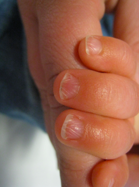
Beau’s lines (transverse ridge) and onychomadesis (nail shedding) common in kids! Often seen in a post-viral setting.
Common culprit = hand foot mouth disease!
Beau’s lines (transverse ridge) and onychomadesis (nail shedding) common in kids! Often seen in a post-viral setting.
Common culprit = hand foot mouth disease!

3/
Congenital malalignment of the great toenails – lateral deviation of the first toenails. More common than you think. Start looking at more toes and you’ll see it! Can improve with time or persist. Risk for nail thickening or ingrown nails.
pc: sciencedirect.com/science/articl…
Congenital malalignment of the great toenails – lateral deviation of the first toenails. More common than you think. Start looking at more toes and you’ll see it! Can improve with time or persist. Risk for nail thickening or ingrown nails.
pc: sciencedirect.com/science/articl…

4/
Baby toes! Congenital hypertrophy of the nail fold describes an overgrowth of the nail fold covering the nail plate. Can treat with topical steroids if inflamed. Will improve by time of that first birthday smash cake!

Baby toes! Congenital hypertrophy of the nail fold describes an overgrowth of the nail fold covering the nail plate. Can treat with topical steroids if inflamed. Will improve by time of that first birthday smash cake!


5/
Chevron nails!
Normal variant…and quite beautiful! Diagonal ridging of the nails most commonly seen in elementary age kids and resolved by adulthood.
pc: onlinelibrary.wiley.com/doi/10.1111/pd…
Chevron nails!
Normal variant…and quite beautiful! Diagonal ridging of the nails most commonly seen in elementary age kids and resolved by adulthood.
pc: onlinelibrary.wiley.com/doi/10.1111/pd…

6/
Punctate leukonychia! Small white spots on the nail plate due to minor trauma to the distal nail matrix. Should grow out with time!
Punctate leukonychia! Small white spots on the nail plate due to minor trauma to the distal nail matrix. Should grow out with time!

7/
Trachyonychia!
Sometimes called “20 nail dystrophy” but does not have to involve all nails! Longitudinal ridging and roughness of the nail plate. Can be idiopathic in childhood or a sign of psoriasis or lichen planus. 50% will self resolve!
pc: journals.lww.com/co-pediatrics/…
Trachyonychia!
Sometimes called “20 nail dystrophy” but does not have to involve all nails! Longitudinal ridging and roughness of the nail plate. Can be idiopathic in childhood or a sign of psoriasis or lichen planus. 50% will self resolve!
pc: journals.lww.com/co-pediatrics/…

8/
Congenital onychodysplasia of the index finger (COIF)!
Tiny nail plate of the second finger. No treatment needed. Classic xray shows bifurcation of the distal phalanx!
#radiology #pedsrads @pedsimaging
pc: doi.org/10.53347/rID-1…
sciencedirect.com/science/articl…

Congenital onychodysplasia of the index finger (COIF)!
Tiny nail plate of the second finger. No treatment needed. Classic xray shows bifurcation of the distal phalanx!
#radiology #pedsrads @pedsimaging
pc: doi.org/10.53347/rID-1…
sciencedirect.com/science/articl…


9/
Pachyonychia congenita, usually presents around 2-3 yo with thickened nails. Spectrum of genetic disorders presenting with thick yellow-brown nails, thick palms and soles, plantar pain.
PC: @pachyonychia
Pachyonychia congenita, usually presents around 2-3 yo with thickened nails. Spectrum of genetic disorders presenting with thick yellow-brown nails, thick palms and soles, plantar pain.
PC: @pachyonychia

10/
Lichen striatus involving the nail! Look for the clues of linear papules leading up to the nail. Can see onycholysis, ridging, splitting, or “pseudo-growth” under the nail plate. Self resolves!
pc: sciencedirect.com/science/articl…
Lichen striatus involving the nail! Look for the clues of linear papules leading up to the nail. Can see onycholysis, ridging, splitting, or “pseudo-growth” under the nail plate. Self resolves!
pc: sciencedirect.com/science/articl…

11/ Melanonychia in kids!
Most often due to congenital nevi or nail matrix nevi. Can look scary! Melanoma in kids is VERY rare, but always feel free to consult a (pediatric) dermatologist for their expert opinion.
Most often due to congenital nevi or nail matrix nevi. Can look scary! Melanoma in kids is VERY rare, but always feel free to consult a (pediatric) dermatologist for their expert opinion.

12/12
In summary, most nail changes in kids are benign. Being able to explain to parents what they are seeing & what to expect will be so appreciated!
Hope all current/future #dermatologists & #pediatricians found this helpful!🙏@Nickles_Melissa for helping prepare this thread!
In summary, most nail changes in kids are benign. Being able to explain to parents what they are seeing & what to expect will be so appreciated!
Hope all current/future #dermatologists & #pediatricians found this helpful!🙏@Nickles_Melissa for helping prepare this thread!
• • •
Missing some Tweet in this thread? You can try to
force a refresh


