“COVID toes” are swollen discolored toes (and fingers) that were seen in areas with high incidence of COVID-19, but the cause is unknown. This new study by @JeffGehlhausen et al shows lack of association between covid toes and SARS-CoV-2 infection. 🧵(1/)
We enrolled 23 pandemic chilblains (PC) patients. While there is an association with community COVID cases (blue line) and PC (red bars), only 2 PC patients had evidence of infection by PCR or antibodies. We wondered if people may have missed the time window for testing +ve. (2/)
PCR testing was difficult to access at the time of initial wave (2020). Thus, we employed two distinct measures of antibodies - ELISA and @serimmune SERA assays - against SARS-CoV-2 S, RBD and N. Only 2 of the 23 patients (who were also PCR +ve) had consistent antiviral Abs. (3/)
However, Ab responses wane over time. So we next tested TCR repertoire of the PC patients using @AdaptiveBiotech’s immunoSEQ T-MAP COVID platform. This assay is more sensitive than Ab assay. It picked up one additional patient, PC #10, as testing pos for antiviral T cells. (4/)
Using an orthogonal approach, we conducted a T cell stimulation assays of PBMCs utilizing a pool of S-protein peptides. This analysis also picked up patient #10 as being positive for T cells against S (consistent with immunoSEQ data). Thus patient #10, 12 and 16 are positive.(5/)
Spike antigen in the toe has been detected by some and suggested as a cause of PC.When we used anti-S Ab for immunohistochemistry (IHC), we saw nice signals for spike in the PC skin (left). However, the same Ab also stained pre-pandemic tissues (right) -> Ab non-specific. (6/)
In fact, we tested a few commercially available antibodies to the S and N and found cross-reactivity to human tissues from the prepandemic era. This is why we need multiple different approaches to ensure what we see in IHC is real. (7/)
So only 3 of 23 pandemic chilblains patients were found to be COVID positive by PCR/Ab/T tests. What accounts for the rest of the patients’ skin lesions? We tested autoantibodies to human exoproteome with REAP with @Aaronmring team and found no increase in PC over control. (8/)
So what can explain pandemic chilblains? Strong mucosal innate immune responses (interferons) that eliminated virus at exposure but caused delayed onset skin inflammation? Seronegative abortive infections by cross-protective T-cells that somehow led to delayed skin rash? (9/)
What immune cell features are most predictive of COVID outcomes? @mkuchroo@JcsHuang Patrick Wong et al used ML algorithm Multiscale PHATE to assign each immune cell type in COVID patients a mortality-likelihood score. Latest from @KrishnaswamyLab 💪🏼 (1/) go.nature.com/3K0QCqi
Based on the flow cytometry data on 54 million cells from COVID 168 patients, the low density granulocytes (neutrophils and eosinophils) were the most enriched cell types in patients who had fatal COVID, followed by inflammatory monocytes and certain B cell subsets. (2/)
In contrast, T cells (most of them; see below), NK cells and dendritic cells were associated with the lowest mortality likelihood scores. They are likely protecting the host from lethal disease. (3/)
Vaccines that reduce infection & disease are needed to combat the pandemic. Here, @tianyangmao@BenIsraelow et al. describe our new mucosal booster strategy, Prime and Spike, to induce such immunity via nasal delivery of unadjuvanted spike vaccine 🧵 (1/)
Current COVID vaccines are given intramuscularly. This induces robust circulating antibodies and systemic T & B cell responses that block viral spread and disease. However, to better block infection, immunity has to be established at mucosal surfaces. (2/) annualreviews.org/doi/10.1146/an…
To elicit mucosal immunity from scratch, live attenuated vaccines are often necessary, due to the need to introduce sufficient antigen and innate immune signals needed for priming via mucosal surfaces. Live vaccines are not safe for immunocompromised. (3/) nature.com/articles/s4157…
So excited to be a part of this important study led by @michelle_monje on how significant longterm neurologic damage can occur after a mild respiratory-only SARS-CoV-2 infection. My own🧵on the findings of this study with relevance to #longCovid (1/)
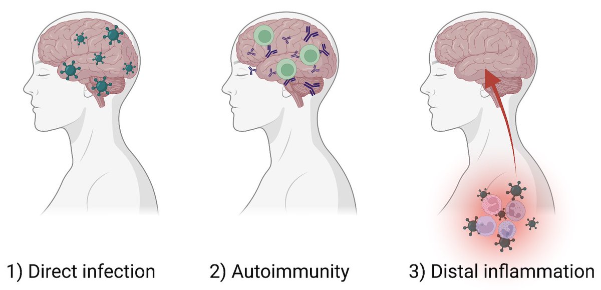
How can a mild respiratory SARS-CoV-2 infection lead to longterm neurological symptoms? Possibilities include 1) direct infection of 🧠, 2) autoimmunity, and 3) inflammatory impact of infection distal to the 🧠. In this study, we focused on 3) 👇🏽 (2/)
To achieve this goal, @peowenlu & @ericsongg used a mouse model developed by @BenIsraelow & @ericsongg in which we can control where the infection happens. Using AAV-hACE2 intratracheally, we can confine the SARS-CoV-2 infection only to the lungs. (3/)
CoronaVac is an inactivated SARS-CoV-2 vaccine approved for use in 48 countries. In collaboration with the Ministry of Health in Dominican Republic, we tested whether CoronaVac (2x) + Pfizer booster induces neutralizing Abs to Delta and Omicron. (1/)
We analyzed plasma samples from 101 participants in Dominican Republic (DR) who received the BNT162b2 booster >4 weeks after the 2x of CoronaVac. We compared them to samples from people at Yale who received 2x of BNT162b2.
⬆️ ⬆️ Ab induced by heterologous prime & boost.(2/)
Next, @carolilucas & @ValterVSM analyzed NAb against ancestral vs. Delta variant. CoronaVac 2x followed by Pfizer mRNA vax booster robustly elevated NAb against both virus types, level similar to 2x Pfizer vaccine. (3/)
This thread is about our new preprint on transposable element called long interspersed nuclear elements (LINE-1/L1). When expressed excessively in the cerebellum, L1 causes ataxia (impaired coordination).
Human genome is occupied in large part by transposable elements or jumping genes. LINE-1 occupies ~20% of our genome, compared to only 1.1% by exons. Some evolutionarily young LINE-1 are still active and are a rare cause of genetic diseases. (2/)
In addition, L1 may even promote the process of aging & age-related diseases in humans. Until recently, research focused on their activity in the germline. Now, their activity in somatic tissues during a lifespan is being studied. Fascinating review👇🏽(3/) nature.com/articles/s4158…
Our new study by @JieunOh9@ericsongg@MiyuMoriyama et al shows that immune priming via intranasal route provides superior protection against heterologous respiratory virus challenge. The key is in inducing local secretory IgA with broader coverage. (1/)
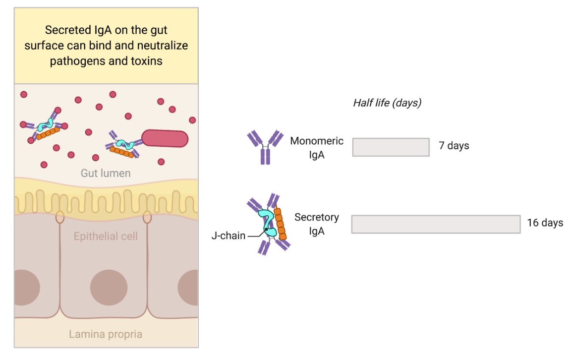
Mucosal surface epithelium expresses polymeric Ig receptor (pIgR), which transports dimeric IgA + J-chain secreted from plasma cells within the tissue, across to the luminal side. IgA dimer + J-Chain + part of pIgR is released as ‘secretory IgA’. Figure by @BioRender. (2/)
The secretory IgA can bind viruses, bacteria, toxin in the lumen of intestine and neutralize them. Advantages of secretory IgA is the extended longevity as well as having 4 Fab instead of 2 Fab (monomeric IgA) to bind to the antigen. Secretory IgA is well studied in the gut. (3/)