1/Mike Tyson once said, “Everyone has a plan until they get punched.”
In honor of all the great hooks and crosses—here is a #tweetorial about mandibular fxs! #FOAMed #medtwitter #Meded #neurorad #HNrad #FOAMrad #Medstudenttwitter @MedTweetorials Image
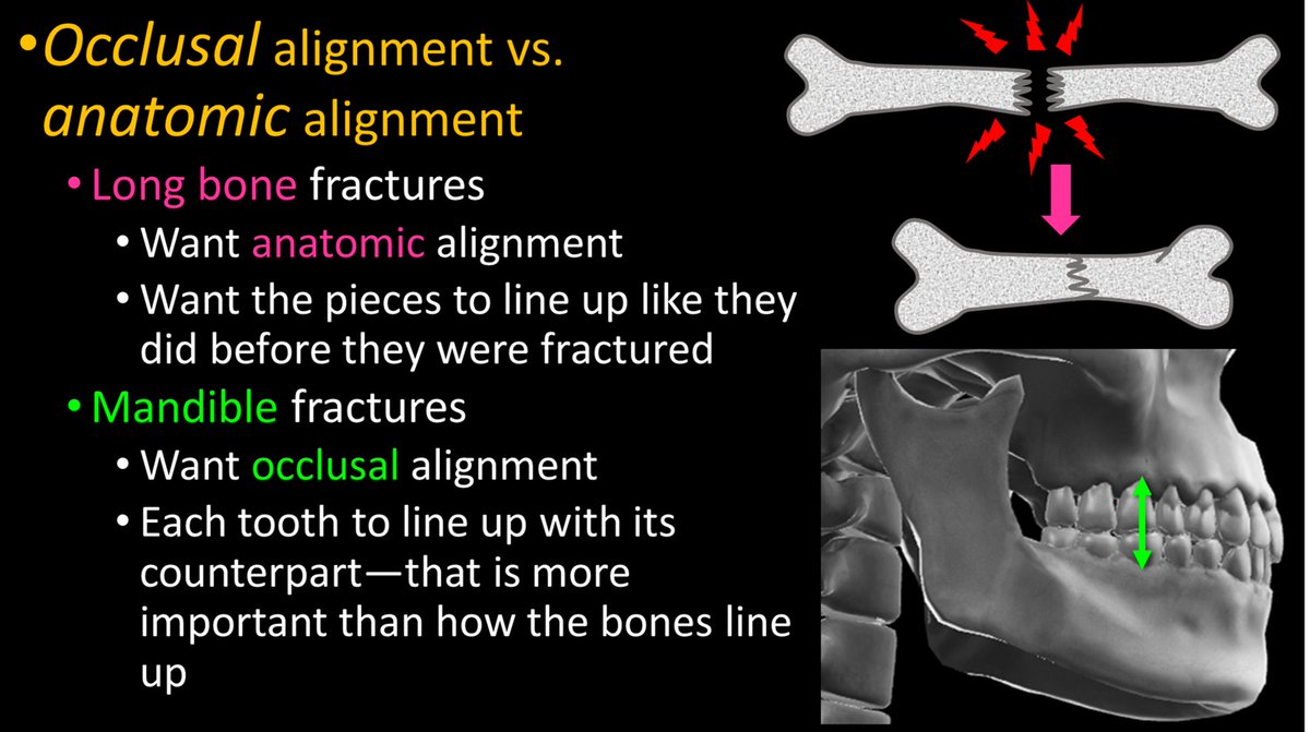
2/A key concept in these fxs is dental occlusion. The jaw is meant to chew. To crush food, teeth need to come as close together as possible—occlusion. Each tooth needs to meet up with its counterpart that fits with it, so no room is left for food—and food will be crushed. Image
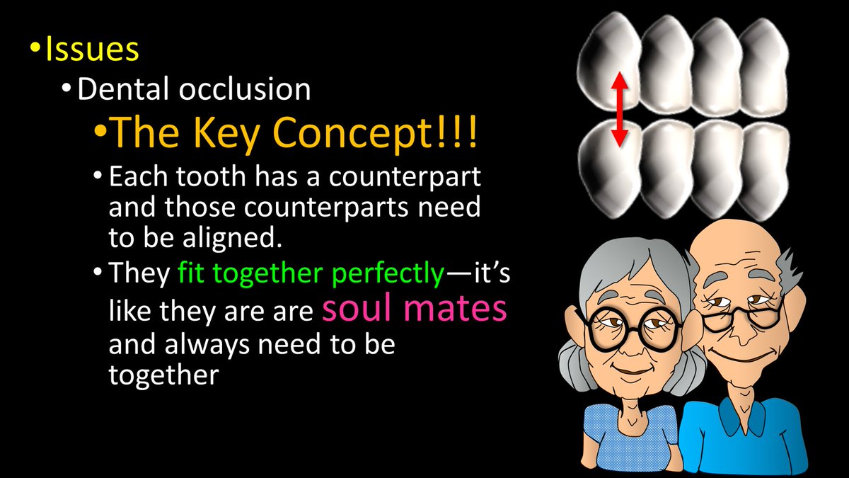
3/Occlusion can be lost w/a fx. The importance of dental occlusion makes mandibular fxs different from other fxs. Usually, we want to fix a bone so that it lines up again. But for the mandible, we want to fix it so the TEETH line up again—so chewing will work. Image
4/Another important concept is multiplicity. It used to be taught that the mandible was a ring w/the skullbase—& you can’t break a ring in one place, so every mandible fx had to have a 2nd one. But this is only true ~50% of time—but it is still worth it to look for the 2nd fx. Image
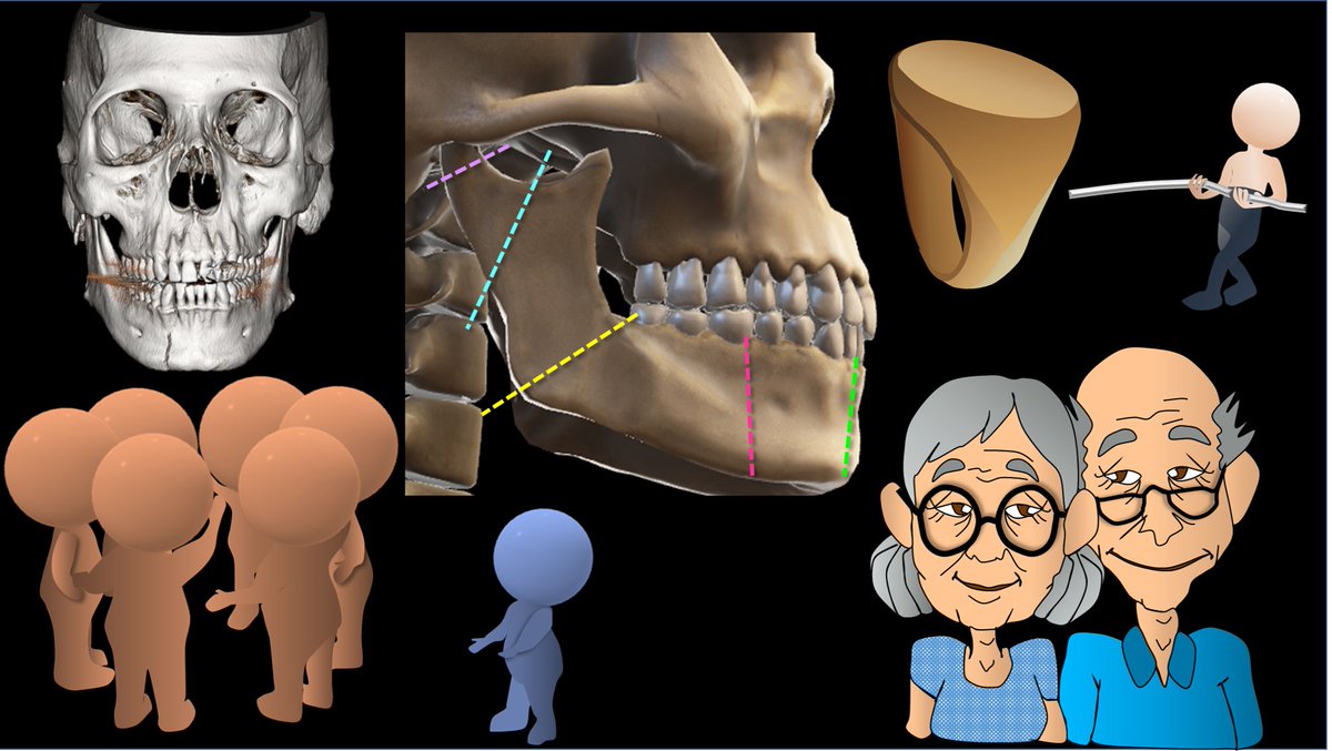
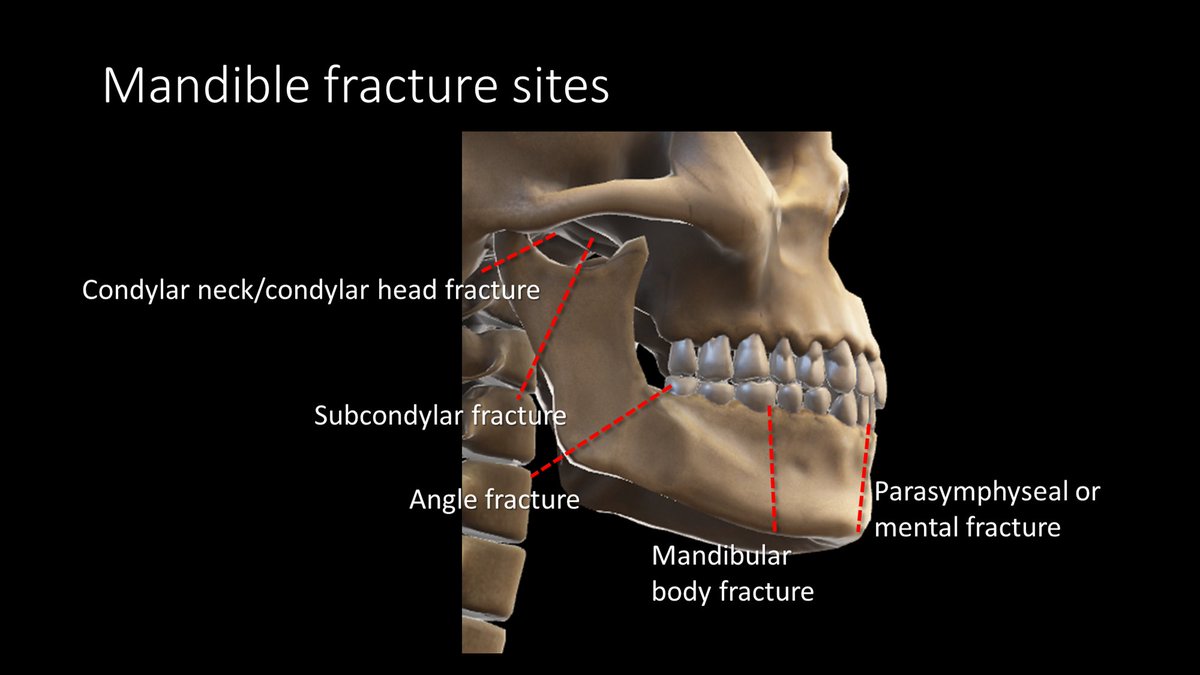
5/Because of the shape of the mandible and the typical forces applied to it, mandibular fractures usually occur at the same few locations. But the names of these fxs are not always intuitive—at least not without a tweetorial to help explain them! 😉 Image
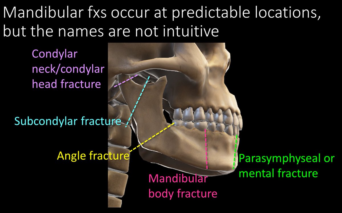
6/Well, perhaps I lied a little. The most common fx does have an intuitive name. An angle fx starts at…wait for it…the angle and extends into socket of the 3rd molar. If you can’t remember an angle fx starts at the mandibular angle, then not even this tweetorial can help you! Image
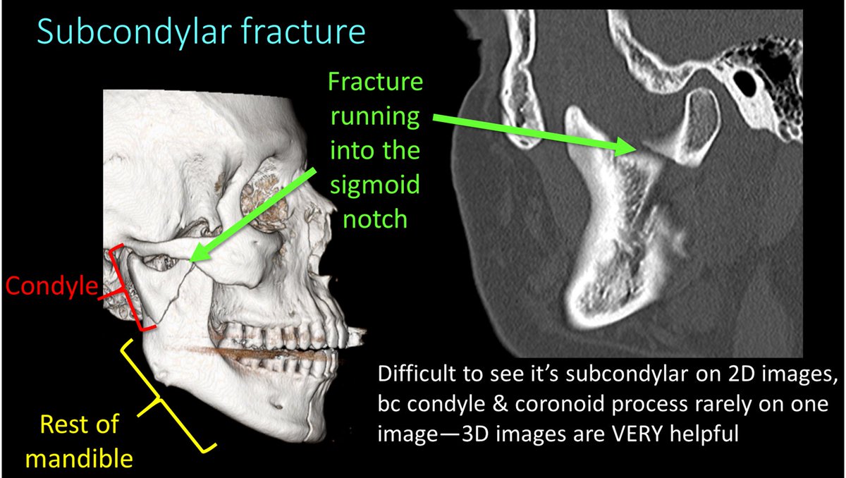
7/A subcondylar fracture starts from the notch between the condyle and coronoid process, called the sigmoid notch and extends into the posterior ramus. Don’t call this a ramus fx bc a ramus fx goes straight horizontally through the ramus! Image
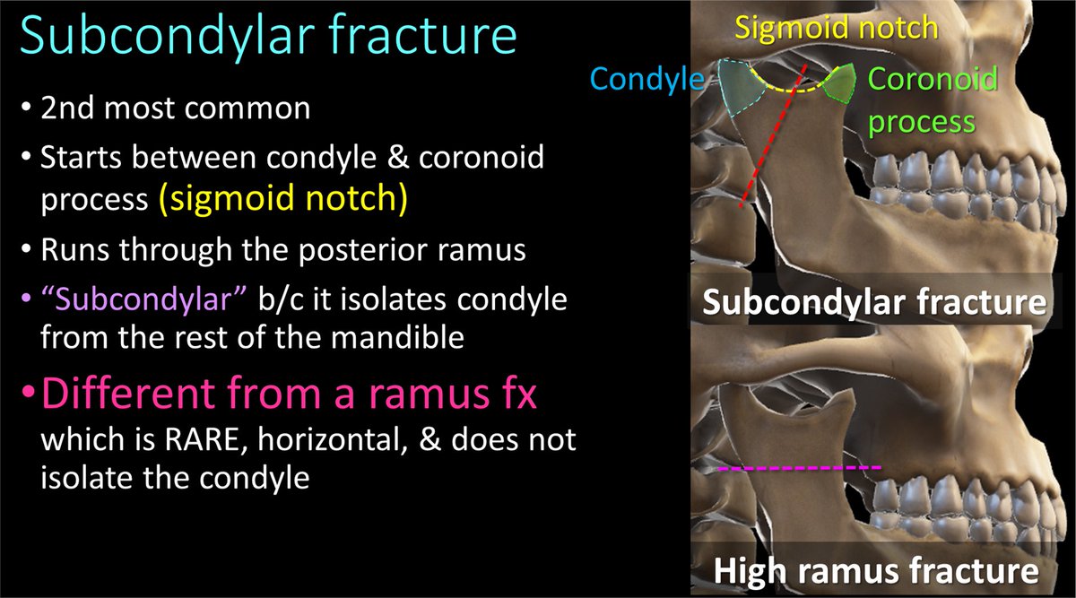
8/Here are examples of subcondylar fxs. A key finding in subcondylar fxs is that it separates the condyle from the rest of the mandible. It can be difficult to see the fx running through the sigmoid notch & ramus on 2D images—3D images can be helpful to see the fx anatomy. Image
9/I remember subcondylar fxs bc they separate the condyle from the rest of the mandible. If you isolate someone, you make them feel SUBpar—so SUBcondylar fxs make the condyle feel subpar bc they separate it from the rest of the mandible! Image
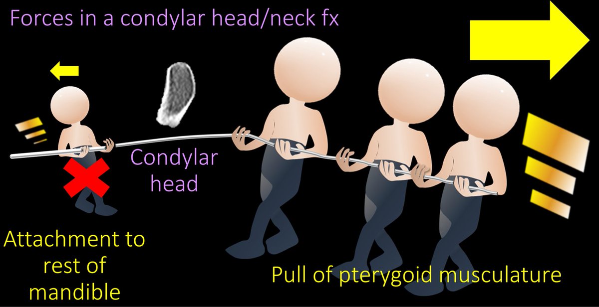
10/Condylar head/neck fxs are easy to remember—they are through, well, the condylar head or neck. These are fx/dislocations. Pterygoid muscles attach to the head & pull medially. So when the head is no longer attached to the mandible, pterygoids are unopposed & pull it medially. Image
11/So if you ever see a condylar head displaced medially, you know it is a condylar head or neck fracture bc this is where the pterygoids are unopposed and pull medially! Image
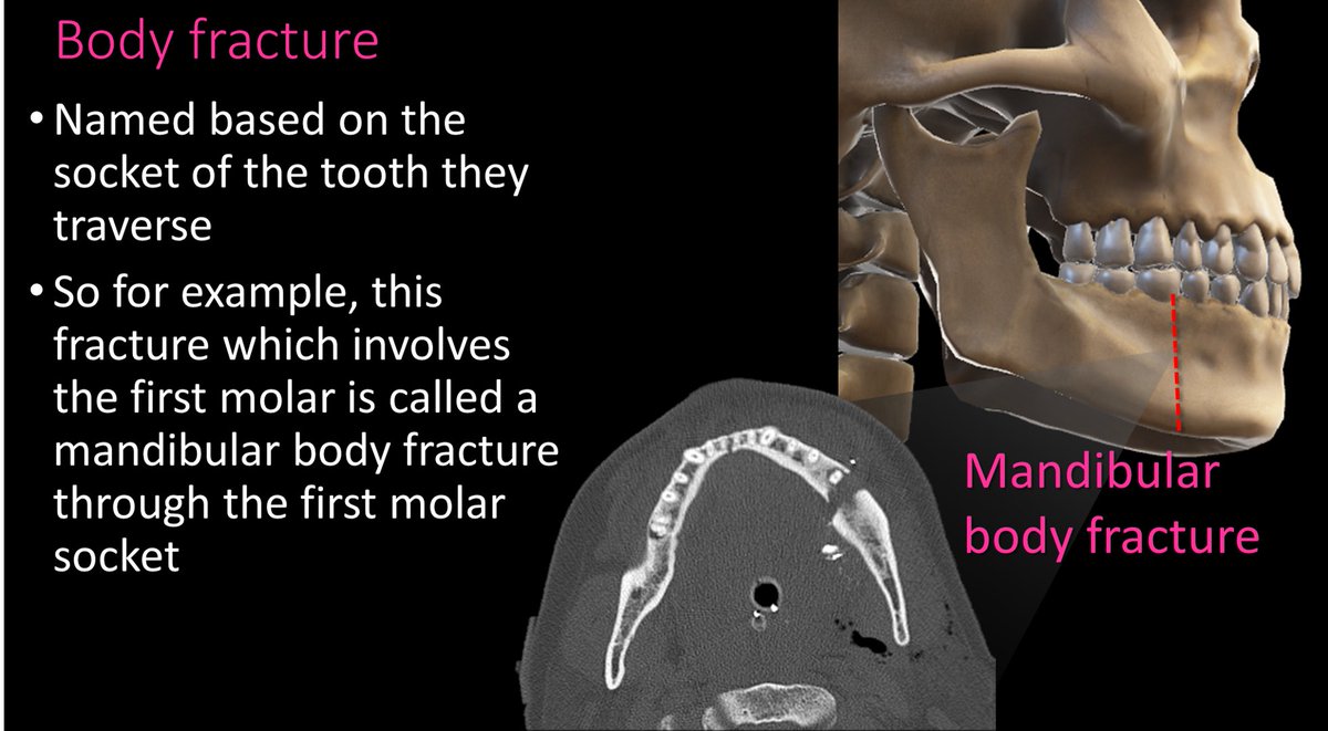
12/Body fxs are through the body of the mandible and are named for the tooth socket that they involve. So you would say “A mandibular body fx through the FILL IN THE BLANK TOOTH socket.” Image
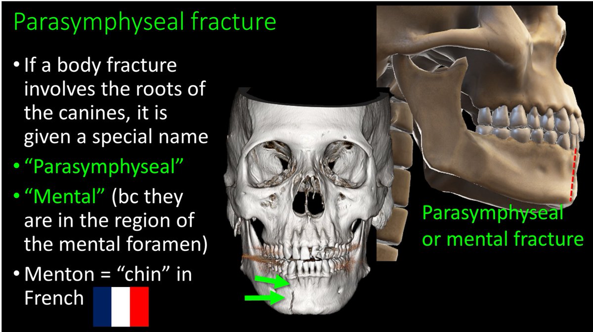
13/Body fxs through the canine are given the special name “parasymphaseal” or “mental” fx bc they are near the mental foramen where the inferior alveolar nerve exits. Menton means “chin” in French, so if they are a body fx anteriorly near the chin, they are “mental” fxs Image
14/So now you know your mandibular fractures & you have a plan even when the patient gets punched! Image

• • •

Missing some Tweet in this thread? You can try to force a refresh
 

Keep Current with Lea Alhilali, MD

Lea Alhilali, MD Profile picture

Stay in touch and get notified when new unrolls are available from this author!

Read all threads

This Thread may be Removed Anytime!

PDF

Twitter may remove this content at anytime! Save it as PDF for later use!

Try unrolling a thread yourself!

how to unroll video
  1. Follow @ThreadReaderApp to mention us!

  2. From a Twitter thread mention us with a keyword "unroll"
@threadreaderapp unroll

Practice here first or read more on our help page!

More from @teachplaygrub

Jun 10
1/ Is the voice box a black box for you?! Let's open the pandora's box of the anatomy of the #larynx together

An introductory #tweetorial on laryngeal #anatomy and the supporting structures. #FOAMed #medtwitter #meded #neurorad #HNrad #FOAMrad #medstudenttwitter @MedTweetorials Image
2/Everyone needs some support. Just like the tongue is supported by the hyoid, the main laryngeal support is the cricoid cartilage. It's like your favorite chair, with all the remotes next to it--not only does the cricoid provide support, it also connects structures in the larynx Image
3/ Although not official, the cricoid is like the real first ring of the trachea. Except it has an odd signet ring shape. The bulbous back part that makes it look like a signet ring provides the important articulation with the arytenoids Image
Read 11 tweets
May 30
1/CSF leaks are controversial Some say they are overdiagnosed, others underdiagnosed. How can you make sure you aren’t under or over diagnosing?
A #tweetorial about a brain MRI score for suspected CSF leaks #medtwitter #FOAMed #radres #neurorad #FOAMrad #csfleak @medtweetorials
2/
With CSF leaks, everyone knows about brain sagging. But this can happen w/other pathologies, ie Chiari 1. Other findings can be seen on brain MRI in CSF leaks. But what are these findings? Are some findings more suggestive than others? Do more findings = ⬆️suspicion?
3/Dobrocky et al. looked at 9 quantitative & 7 qualitative signs seen on brain MRI in CSF leaks to see which are most important. Depending on type & # of findings, they developed a score to indicate what level of suspicion you should have for a leak.
pubmed.ncbi.nlm.nih.gov/30776059/
Read 12 tweets
May 20
1/
Everyone loves those pretty colored fMRI pictures with the blobs. But what do they mean? How do they make them?
A #tweetorial called “F— that: Understanding fMRI”. #FOAMed #medtwitter #Medstudenttwitter #neurotwitter #FOAMrad #neuroradiology #radiology
2/
fMRI is based on a principle called “neurovascular coupling.” This is the principle if there is increased neuronal activity in a region, there will be increased blood flow to that region to meet the increased demand
3/
Think of it like a baby crying because it is hungry—parents immediately rush to feed it. The increased oxygen demand of the neurons immediately brings increased fuel to feed it.
Read 17 tweets
May 18
1/
“You don’t get points for having your needle in the right place if you don’t get a diagnosis.” When we biopsy the skullbase we work to get a diagnosis.
A sort of #tweetorial but more like a 🧵about our skullbase biopsy system. #FOAMed #medtwitter #neurosurgery #neurotwitter Image
2/
Unless the lesion is difficult to diagnosis w/FNA (ie, schwannoma), we begin by FNA w/an 18g draw needle & a 22g Quincke needle. We do not aspirate, b/c the skullbase is very vascular, & too much blood will be drawn up, making it difficult to tell if the sample is diagnostic. Image
3/
However, if we are not getting a diagnosis with FNA, we will move to a core. If it is a deep lesion, we will use the Biopince system, beginning with a 17g, 7 cm introducer. This is an example of IgG4 disease of the trigeminal nerve that failed FNA and required a core Image
Read 7 tweets
May 16
1/
Why is cranial nerve 6 uniquely affected by⬆️ intracranial pressure? Why is it special? A common question after the CN6 tweetorial.
Here is a maybe #tweetorial, but maybe a🧵about why CN6 is alone affected by ⬆️ pressure. #FOAMed #medtwitter #Medstudenttwitter #neurotwitter Image
2/
Think of the intracranial CSF space like a balloon, distended by CSF instead of air. Cranial nerves begin inside the balloon, and then they exit as they begin their extradural portion Image
3/
Most cranial nerves move immediately away from the CSF space after they exit—usually going out through their respective foramina. However, CN6 uniquely runs along the outside of the “balloon” in Dorello canal Image
Read 7 tweets
May 13
1/
It is rare a patient tells you where their pathology is—but they do when they tell you they have a cranial nerve palsy—you just have to know where to look!
A #tweetorial about the Six Syndromes of the Sixth Cranial Nerve. #FOAMed #medtwitter #Medstudenttwitter @medtweetorials
2/
To understand the six syndromes, you have to know the anatomy of the 6th nerve. It starts in the brainstem, at the facial colliculus—what looks like the butt of the brainstem. It then travels anteriorly through the brainstem to exit out the ventral surface of the pons.
3/
It exits the brainstem at the inferior aspect of the pons—under what looks like pot belly of the pons and then travels anteriorly in the subarachnoid space towards the clivus
Read 17 tweets

Did Thread Reader help you today?

Support us! We are indie developers!


This site is made by just two indie developers on a laptop doing marketing, support and development! Read more about the story.

Become a Premium Member ($3/month or $30/year) and get exclusive features!

Become Premium

Don't want to be a Premium member but still want to support us?

Make a small donation by buying us coffee ($5) or help with server cost ($10)

Donate via Paypal

Or Donate anonymously using crypto!

Ethereum

0xfe58350B80634f60Fa6Dc149a72b4DFbc17D341E copy

Bitcoin

3ATGMxNzCUFzxpMCHL5sWSt4DVtS8UqXpi copy

Thank you for your support!

Follow Us on Twitter!

:(