2/First, a rule of thumb—or rather a rule of elbow! You have 10 fingers. If you divide that in half, you get 5. Similarly, if you divide your arm in half at the elbow, you get 5--C5 that is! C5 radiates towards the elbow. So if it radiates below this, it is > C5 & above is < C5
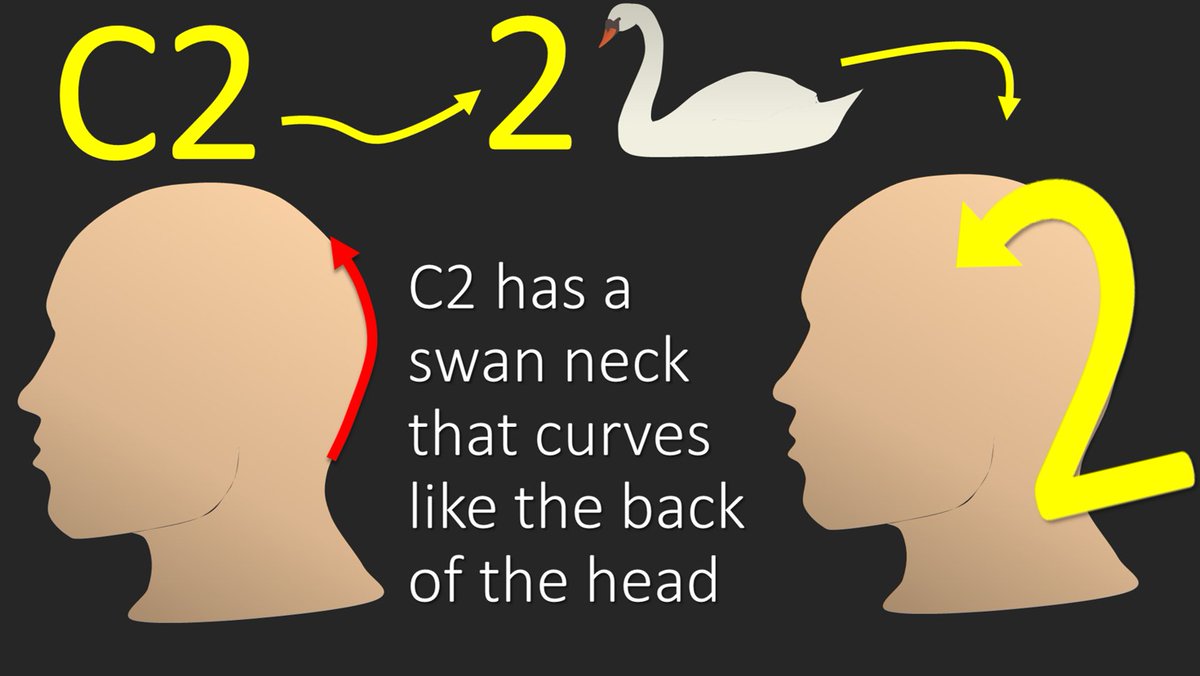
3/So let’s start with C2. C2 predominantly radiates along the dorsal aspect of the scalp, as it supplies the greater occipital nerve. I remember this bc the number 2 has a swan like neck that mimics the contour of the back of the head and its distribution
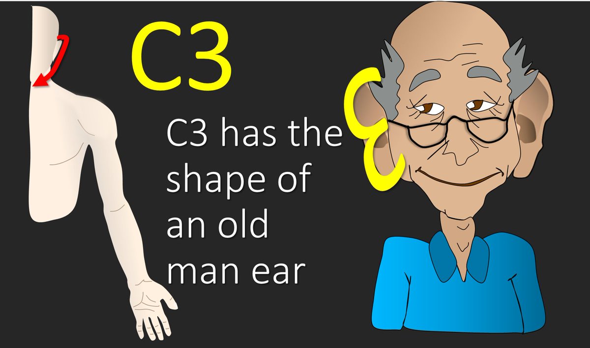
4/Next is C3. C3 starts radiating right around the ear and circles around to the front of the neck, as it is a contributor to the great auricular nerve and anterior cutaneous nerve of the neck. I remember how it starts bc the number 3 looks like those big grandpa ears.
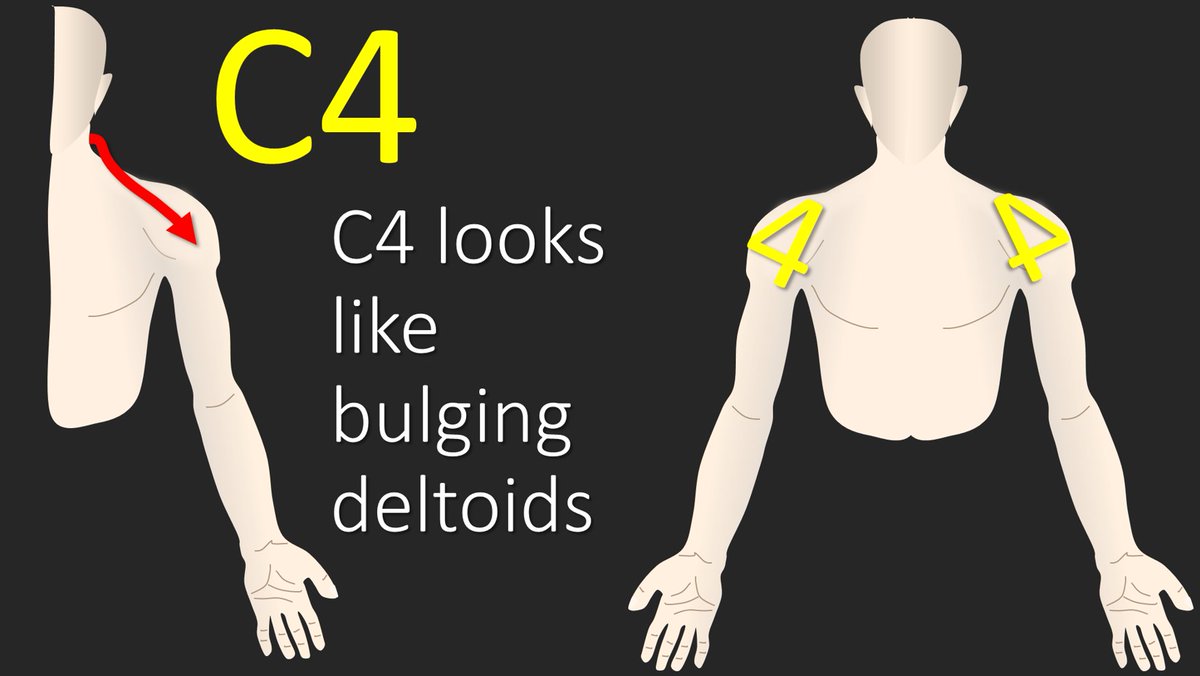
5/C4 radiates along the suprclavicular fossa, as it helps innervate the supraclavicular nerve, and goes towards the deltoid. I remember this because the triangular part of the number 4 looks the bulging deltoids that you always see on TV, but never in real life 😂
6/I remember C5 using that general rule that half of the number of fingers (5) equals halfway down the arm (elbow). C5 radiates towards the elbow.
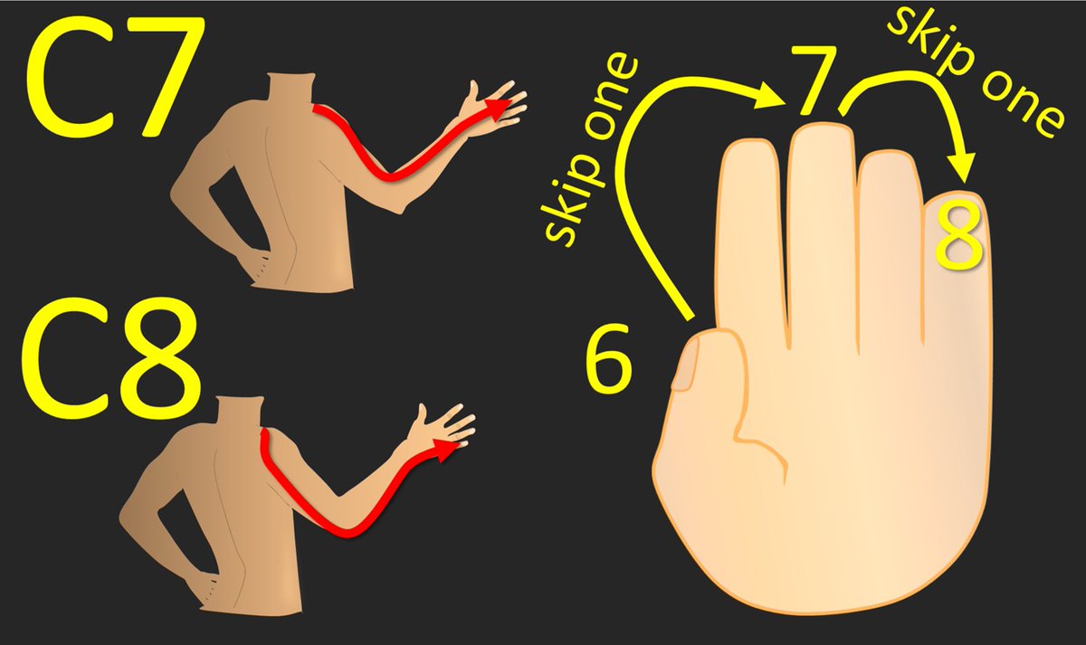
7/C6 radiates to the thumb. I remember this because when you count to 6, you use up all the fingers on one hand and then end on the thumb of the next hand. So you end on a thumb when you count to 6 and C6 radiates to the thumb
8/We only have two cervical nerves left (C7, C8) for the 4 fingers left. So we will just use every other finger. So starting with C6 at the thumb—skip a finger gets you to the middle finger, that’s C7, skip the next finger gets you to the pinky, and that’s C8!
9/Now you know where in the c-spine to look when a patient has pain radiating from the neck—but remember there are subtleties to this & these are general guidelines to help you. Hopefully, remembering cervical radicular pain distributions will no longer be a pain in the neck!
• • •
Missing some Tweet in this thread? You can try to
force a refresh
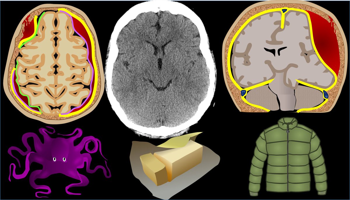
2/In bread & butter neuroimaging—CT is the bread—maybe a little bland, not super exciting—but necessary & you can get a lot of nutrition out of it. MRI is like the butter—everyone loves it, it makes everything better, & it packs a lot of calories. Today, we start w/the bread!
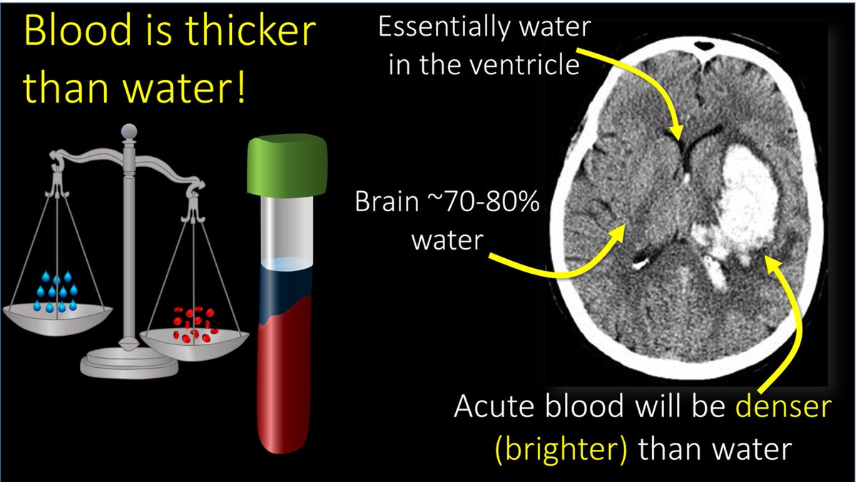
3/The most important thing to look for on a head CT is blood. Blood is Bright on a head CT—both start w/B. Blood is bright bc for all it’s Nobel prizes, all CT is is a density measurement—and blood is denser (thicker) than water and denser things are brighter on CT
2/Let's use this french fry illustration I used for my varices🧵
I use the curve function to create shapes. It has the most control for drawing--the freeform function causes uneven lines.
After drawing the shape, I use the shape fill function to fill it in it w/the color I need
3/ 3D effects begin w/the gradient fill. I use the gradient fill that powerpoint offers for my selected color--it knows how to chose them so that the gradient is smooth
2/Pt with history only of diabetes came in with altered mental status. There were these super bright round balls in their ventricles. It looked like a snowman massacre happened, with big round snowballs in the ventricles
3/On MRI, these had intrinsic T1 hyperintensity w/chemical shift artifact--they looked like boba tea pearls floating in the ventricles. Typically, we think of chemical shift w/fat--but these were not fat on the CT!! They did not enhance & otherwise, brain was unremarkable for age
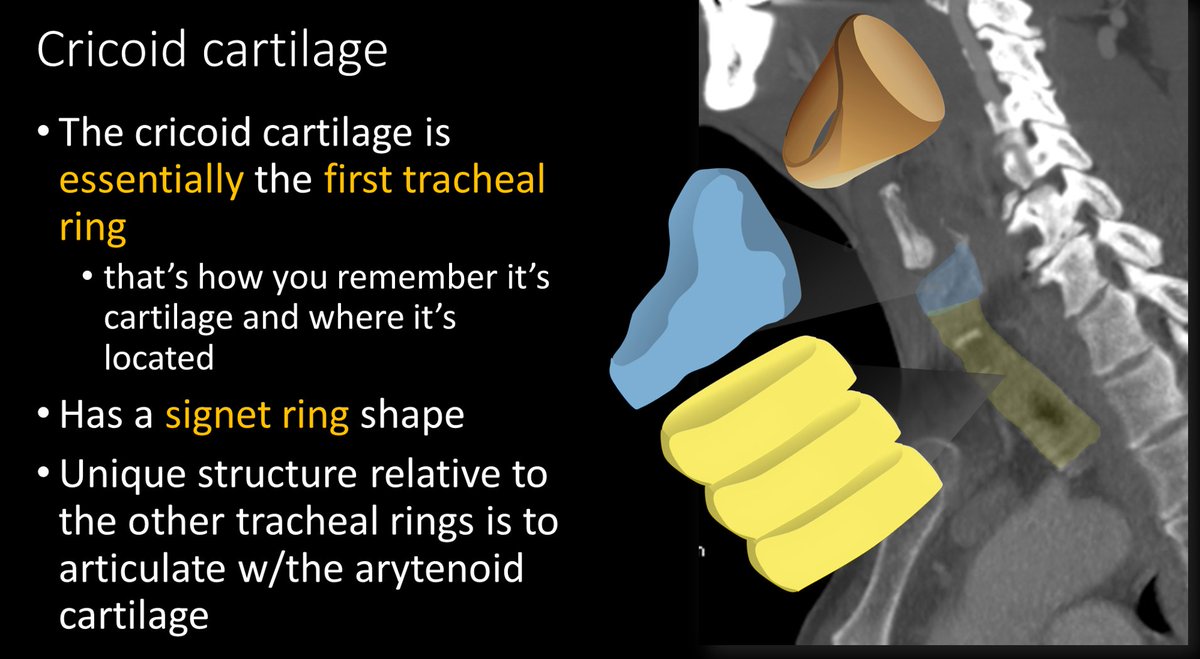
2/Everyone needs some support. Just like the tongue is supported by the hyoid, the main laryngeal support is the cricoid cartilage. It's like your favorite chair, with all the remotes next to it--not only does the cricoid provide support, it also connects structures in the larynx
3/ Although not official, the cricoid is like the real first ring of the trachea. Except it has an odd signet ring shape. The bulbous back part that makes it look like a signet ring provides the important articulation with the arytenoids
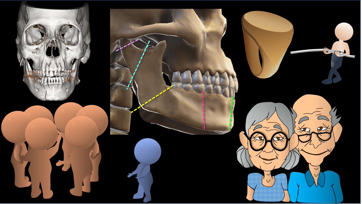
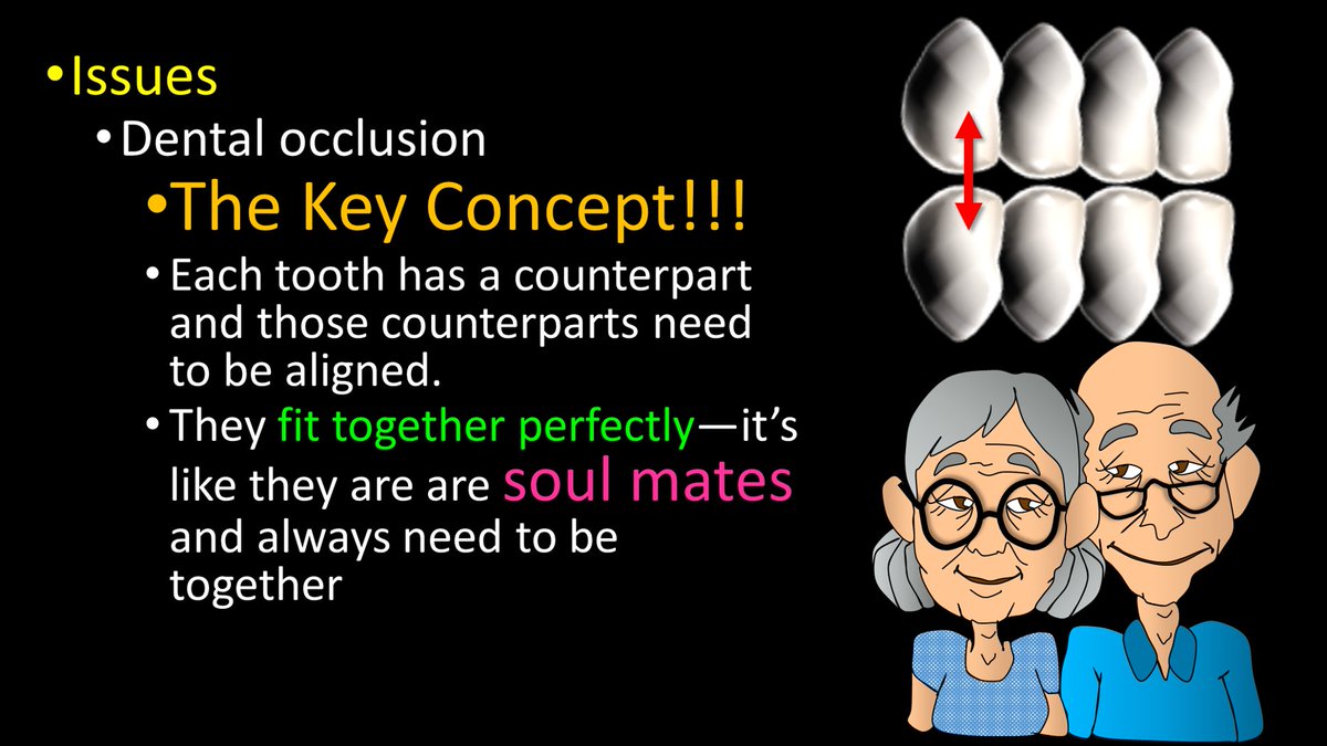
2/A key concept in these fxs is dental occlusion. The jaw is meant to chew. To crush food, teeth need to come as close together as possible—occlusion. Each tooth needs to meet up with its counterpart that fits with it, so no room is left for food—and food will be crushed.
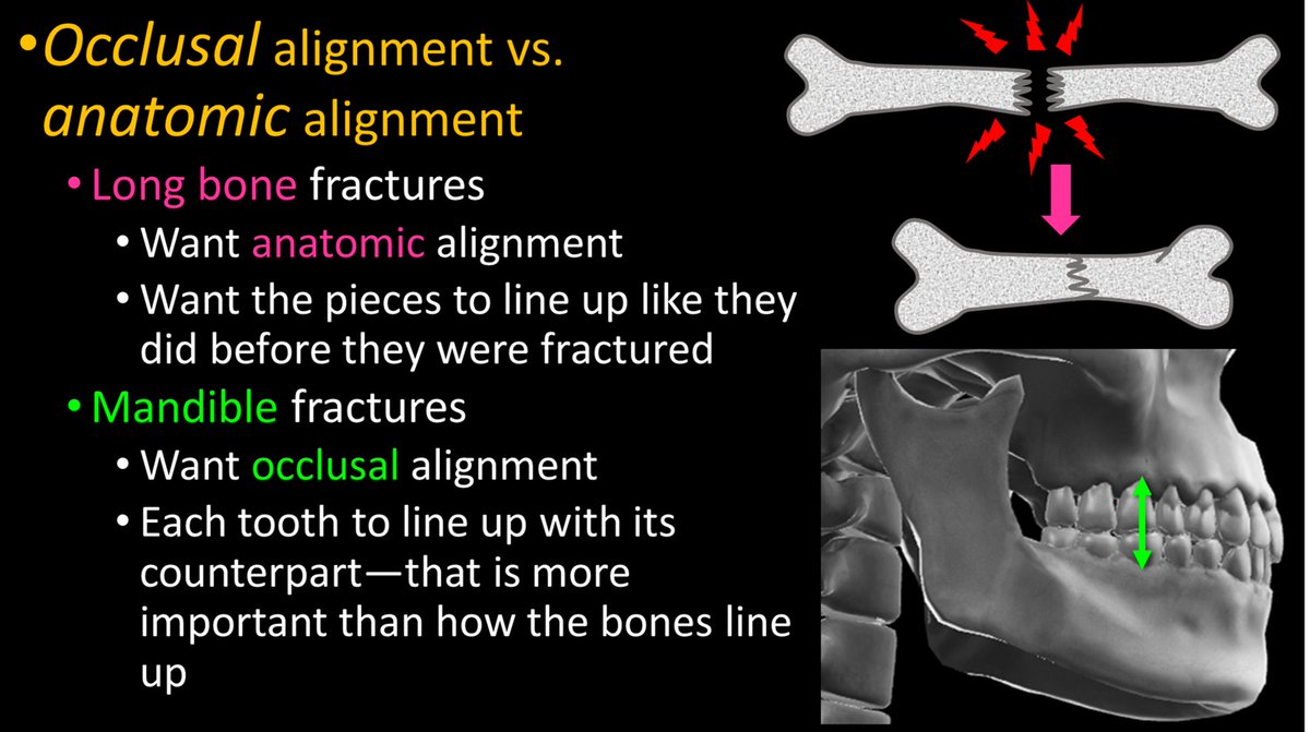
3/Occlusion can be lost w/a fx. The importance of dental occlusion makes mandibular fxs different from other fxs. Usually, we want to fix a bone so that it lines up again. But for the mandible, we want to fix it so the TEETH line up again—so chewing will work.
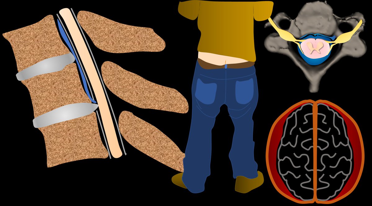
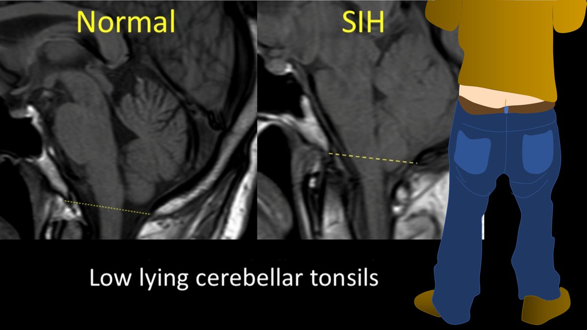
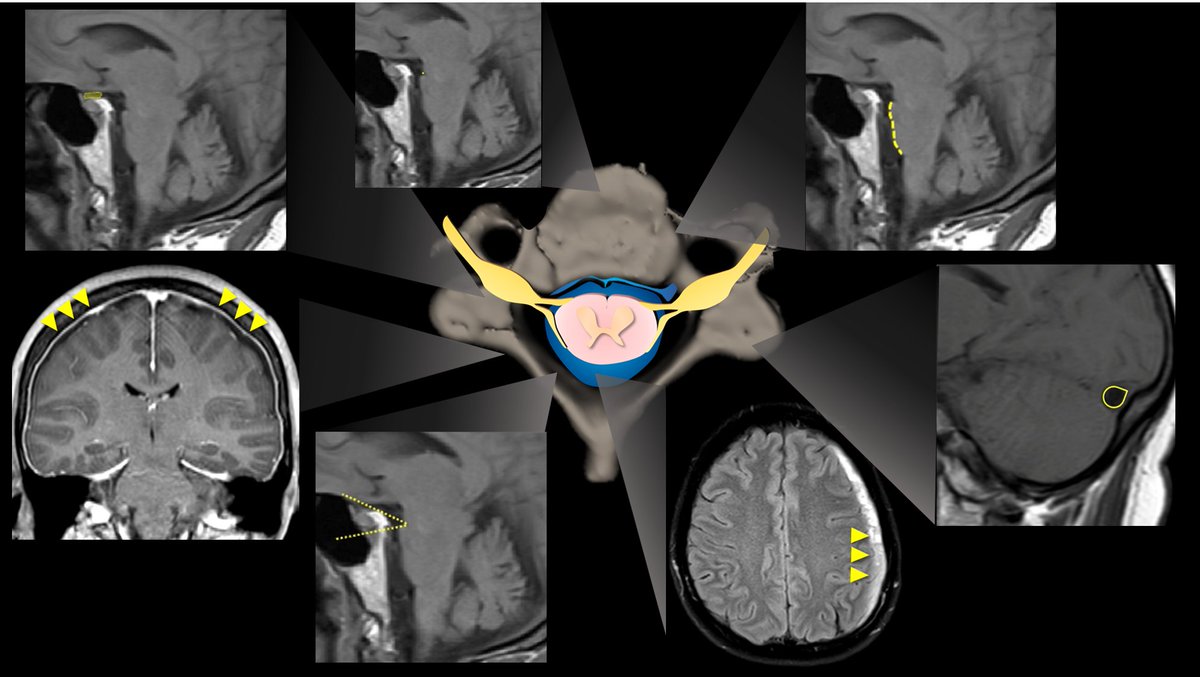
2/ With CSF leaks, everyone knows about brain sagging. But this can happen w/other pathologies, ie Chiari 1. Other findings can be seen on brain MRI in CSF leaks. But what are these findings? Are some findings more suggestive than others? Do more findings = ⬆️suspicion?
3/Dobrocky et al. looked at 9 quantitative & 7 qualitative signs seen on brain MRI in CSF leaks to see which are most important. Depending on type & # of findings, they developed a score to indicate what level of suspicion you should have for a leak. pubmed.ncbi.nlm.nih.gov/30776059/