
1/Hello all,Good morning! Happy independence week🇮🇳🎉🎊
Got a beautiful chest teaching case 👇🏻Xray ▶️CT ▶️HPE
🛑27 year old female came with h/o chest discomfort, breathing difficulty, cough x 1 week
Lets breakdown it✍️📝 🔥
#radres #chestrad #Pulmonology #MedTwitter #FOAMrad
Got a beautiful chest teaching case 👇🏻Xray ▶️CT ▶️HPE
🛑27 year old female came with h/o chest discomfort, breathing difficulty, cough x 1 week
Lets breakdown it✍️📝 🔥
#radres #chestrad #Pulmonology #MedTwitter #FOAMrad

2/ Frontal CXR shows homogenous opacity in R.mid & lower zone in paravertebral region
❇️It has well defined superior,inferior&lateral borders, medial border is merged with mediastinum(incomplete border sign)
♉️Opacity has Broad base contact with mediastinum s/o mediastinal origin
❇️It has well defined superior,inferior&lateral borders, medial border is merged with mediastinum(incomplete border sign)
♉️Opacity has Broad base contact with mediastinum s/o mediastinal origin

3/
❇️Lung lesion will be covered by lung margins all side(not always) & form an acute angle with meds
♉️Normal silhouette of right heart border is seen (neg silhouette sign) s/o lesion not in ant med
🉑Hilar vessels are seen coursing through the opacity (Hilum overlay sign +ve)
❇️Lung lesion will be covered by lung margins all side(not always) & form an acute angle with meds
♉️Normal silhouette of right heart border is seen (neg silhouette sign) s/o lesion not in ant med
🉑Hilar vessels are seen coursing through the opacity (Hilum overlay sign +ve)


4/
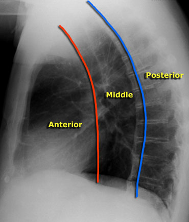
♓️Lateral border of the opacity is >2cm from the hilar vessels¬ converging towards the vessels (Hilum convergence sign-ve) s/o the lesion is not in middle med
❇️Inferior border of the opacity extend below R.diaphrgm s/o lesion in post med as ant med doesn’t extend below it
♓️Lateral border of the opacity is >2cm from the hilar vessels¬ converging towards the vessels (Hilum convergence sign-ve) s/o the lesion is not in middle med
❇️Inferior border of the opacity extend below R.diaphrgm s/o lesion in post med as ant med doesn’t extend below it

5/
❇️No cavitation/Ca++/air fluid level/rib erosion/fracture
♒️The above features suggests that the lesion is of mediastinal origin & most likely in post mediastinum
🛑Lateral xray confirms the lesion is in posterior mediastinum
🉑What are the DD for posterior mediastinal lesion?
❇️No cavitation/Ca++/air fluid level/rib erosion/fracture
♒️The above features suggests that the lesion is of mediastinal origin & most likely in post mediastinum
🛑Lateral xray confirms the lesion is in posterior mediastinum
🉑What are the DD for posterior mediastinal lesion?

Please add your differentials and guess the diagnosis of above case. I will post the CT images & Final diagnosis later on. Thank you
#radres #chestrad #Pulmonology #MedTwitter #FOAMrad #USMLE #NEETPG2022 #MedEd #radiology #medicine
#radres #chestrad #Pulmonology #MedTwitter #FOAMrad #USMLE #NEETPG2022 #MedEd #radiology #medicine
@RadiologyVibes @_the_SRT @thoracicrad @radiologistpage @TLHM_MD @CasesCookyJar @RCC_Editor @AOCRNews @Dx_imaging @NIHRadiology @drdevrad @UCSFChest @nverma21 @ARRS_Radiology @RSNA @Radiopaedia @STS_CTsurgery @APTAcvp @dr_veeprakash
• • •
Missing some Tweet in this thread? You can try to
force a refresh


