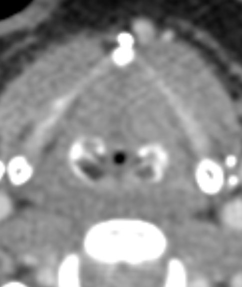
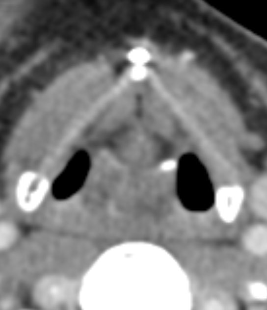
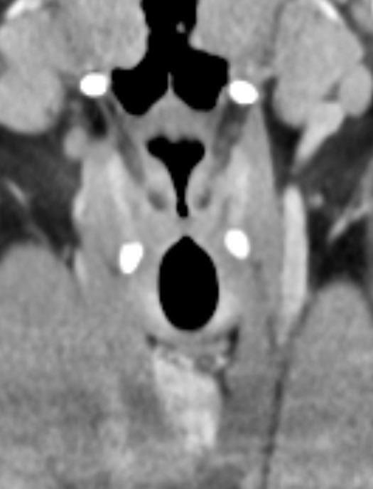
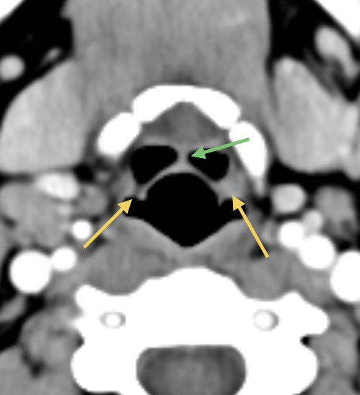
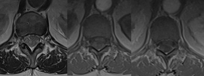
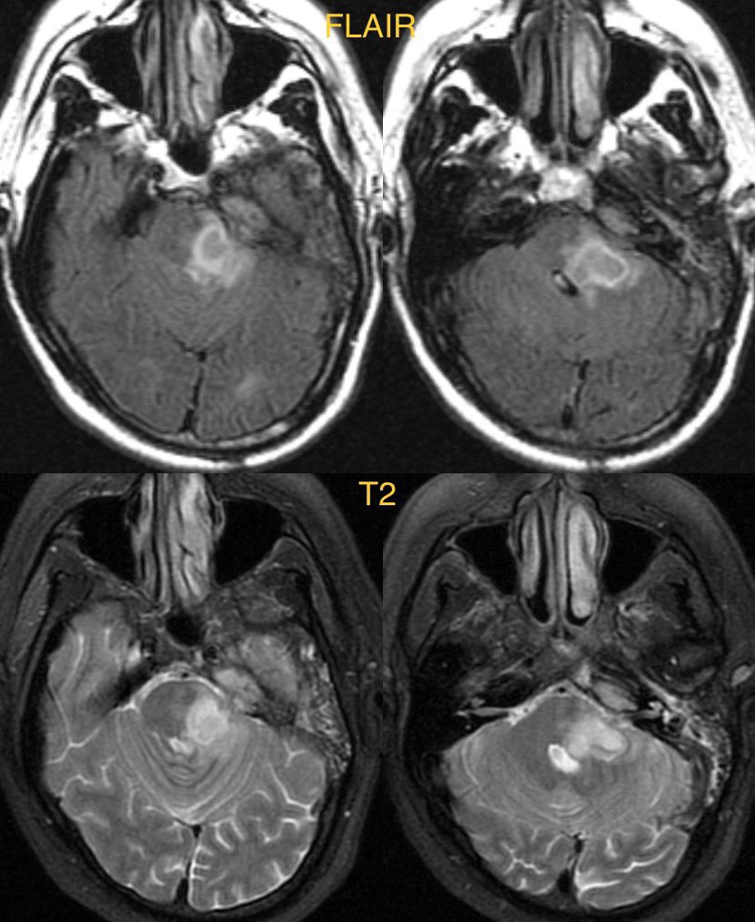
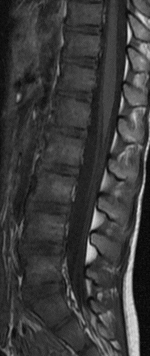
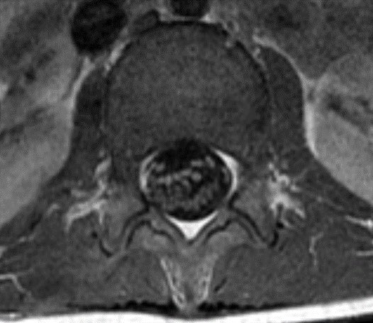
Child presents with weakness. MR shows enhancement of the pial surface of the conus and ventral cauda equina nerve roots.
#radtwitter #MedTwitter #radres #futureradres #Pediatrics #Neurology @TheASNR @The_ASPNR @AlbanyMedRadRes
#radtwitter #MedTwitter #radres #futureradres #Pediatrics #Neurology @TheASNR @The_ASPNR @AlbanyMedRadRes

Differential diagnosis:
Leptomeningeal carcinomatosis
Lymphoma
Arachnoiditis (all causes)
Guillain-barre
Neurosarcoidosis
Leptomeningeal carcinomatosis
Lymphoma
Arachnoiditis (all causes)
Guillain-barre
Neurosarcoidosis
Diagnosis: Guillain-Barré syndrome
These are the typical imaging features for GBS. Contrast is absolutely necessary.
There was no history to suggest systemic sarcoidosis, malignancy, or recent procedure (risk factor for spinal meningitis/arachnoiditis)
These are the typical imaging features for GBS. Contrast is absolutely necessary.
There was no history to suggest systemic sarcoidosis, malignancy, or recent procedure (risk factor for spinal meningitis/arachnoiditis)
• • •
Missing some Tweet in this thread? You can try to
force a refresh