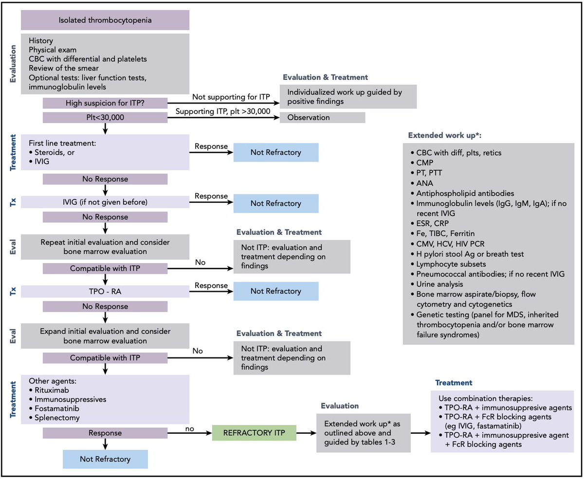
Saw a patient in clinic w/ (likely) refractory ITP and luckily there was an excellent @BloodJournal article by Miltiadous and colleagues published earlier this month! I want to highlight some of the main learning points I took away #medtwitter#hemetwitterashpublications.org/blood/article/…
# 1: Make sure that the patient truly has primary, refractory ITP! If a patient is not responding as expected with primary ITP-directed therapies, it bears consideration to do a more extensive workup looking for alternative diagnoses
#2: If a patient is not responding as expected to treatment for ITP, have a low threshold to pursue a bone marrow biopsy. A number of bone marrow failure states can present with isolated thrombocytopenia
#3: If a patient truly has refractory, primary ITP then combination therapies should be considered. The flowchart nicely outlines workup and treatments, the evidence behind different combinations is covered in the article itself
In summary, the more ITP I see the more I realize how many patients may not have it. Therefore I have a low threshold to pursue additional workup and re-examine the original dx if patients aren't responding as expected. Would love to get additional pearls from #medtwitter
A couple weeks ago I was caring for a patient on the heme-onc service that developed progressive L-sided neck/face swelling over 3-4 hours! He ended up having suppurative parotitis, a diagnosis I have not seen very much in practice. Here are some takeaways #MedTwitter
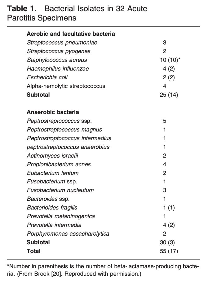
Risk fxrs for suppurative parotitis include poor PO intake, poor dentition/recent dental work, oropharyngeal malignancies, & intubation (all via impaired stimulation/drainage of salivary ducts). The most common offending bugs are Staph Aureus & anaerobes (pubmed.ncbi.nlm.nih.gov/12544218/)
Both US and CT can be used as imaging modalities in the workup of suppurative parotitis. US can identify a potential obstructing stone more easily while CT is more sensitive for ruling out an abscess. Another diagnostic hint is elevated amylase w/ normal lipase
Let's talk about cytopenias in cirrhosis. Many patients with cirrhosis will have at least one cytopenia (sometimes pancytopenia) attributable to their liver disease, let's talk about some of the mechanisms! #medtwitter#hemetwitter
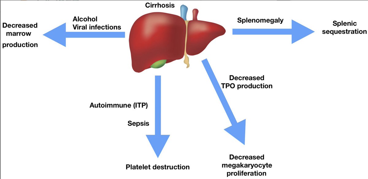
First up, thrombocytopenia! The potential mechanisms are laid out in the image below (aasldpubs.onlinelibrary.wiley.com/doi/full/10.10…):
- decreased thrombopoietin production
- sequestration
- alcohol/viral causes suppressing marrow
- peripheral destruction
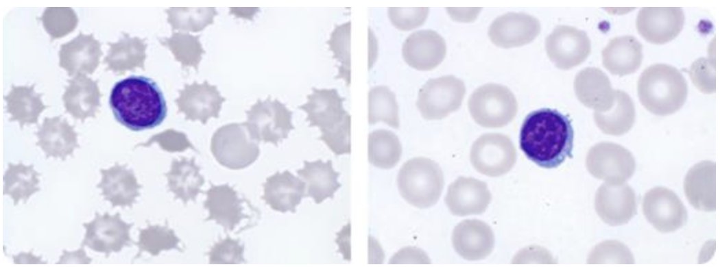
Next up, anemia! The ddx should include the causes we think of in all anemia patients (nutritional, GI losses, hemolysis etc...) but what are some liver-specific etiologies? Spur-cell anemia (ashpublications.org/blood/article/…) typically only reverses w/ transplant
Drs. Gotlib and Reiter put an an excellent review discussing myeloid neoplasms with hypereosinophilia (ashpublications.org/blood/article-…), these are some of the salient points for all IM folks to take away
The first is knowing that the terms hypereosinophilia and hypereosinophilic syndrome (HES) carry specific meaning! HES meets the definition for hypereosinophilia + there is organ damage 2/2 hypereosinophilia
Based on the WHO guidelines, the following values constitute polycythemia:
In men: Hgb >16.5g/dL or Hct > 49%
In women: Hgb > 16 or Hct > 48%
Polycythemia should be confirmed by a subsequent CBC. Below is an image of the ddx of polycythemia (Keohane et al 2013)
Our patient's polycythemia has been confirmed! What to do next? Make sure to ask questions related to hyperviscosity (eg chest pain, dyspnea, headache, vision changes, confusion), thrombosis/bleeding hx, and PV-related symptoms (pruritus after baths, erythromelalgia, B-symptoms)
#Tweetorial Just finished 4 weeks on the leukemia service at @UCCancerCenter. We saw a number of patients present with a new diagnosis of acute leukemia and hyperleukocytosis, our index of concern for the development of leukostasis was dependent on if they had AML or ALL.
In 1982 Lichtman et al wrote about hyperleukocytic leukemias. The leukocrit (packed WBC volume) was described in AML, ALL, and CLL. Due to the larger mean cell volume of myeloblasts, a higher leukocrit was observed in AML compared to ALL and CLL.
The pathophysiology of leukostasis is thought to be driven in part by blood viscosity. A higher leukocrit and hematocrit correlates with a higher blood viscosity.