Wetread Case 40
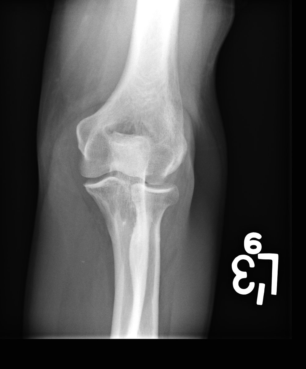
Hx: elbow swelling.
Diagnosis(es)?
Clinician inquires about aspiration. Thoughts?
Please answer with professionally appropriate gif responses only. No spoilers. wetread.org #WetreadCow #FOAMrad #FOAMed #radres #errad #mskrad #orthorad

Hx: elbow swelling.
Diagnosis(es)?
Clinician inquires about aspiration. Thoughts?
Please answer with professionally appropriate gif responses only. No spoilers. wetread.org #WetreadCow #FOAMrad #FOAMed #radres #errad #mskrad #orthorad


Wetread Case 40 Answer
Answer: Olecranon Bursitis secondary to Gout
We have focal mass-like soft tissue density posterior to the olecranon with o/w minimal soft tissue edema. Add on some soft tissue Ca+ and this is classic picture of gouty olecranon bursitis
Answer: Olecranon Bursitis secondary to Gout
We have focal mass-like soft tissue density posterior to the olecranon with o/w minimal soft tissue edema. Add on some soft tissue Ca+ and this is classic picture of gouty olecranon bursitis
Etiologies for olecranon bursitis:
-trauma
-repeated or continued pressure/irritation (aka “student’s", “baker’s” or Popeye elbow)
-infection
-inflammation (classically gout, although RA and CPPD as well)
-trauma
-repeated or continued pressure/irritation (aka “student’s", “baker’s” or Popeye elbow)
-infection
-inflammation (classically gout, although RA and CPPD as well)
In this case we also see soft tissue Ca++ which we wouldn’t expect to see in trauma or overuse but can be seen with gout. Bony changes are not very apparent b/c it usually takes 10+ years for this to occur.
Gout:
-Articular/periarticular deposition of monosodium urate crystals (birefringent & poorly dissolve in synovial fluid)
-middle aged M>>F
-due to hyperuricemia
-undersecrete: CRF, HyperPTH, alcohol, drugs
-overproduce: hemolysis,myeloprolif dz
-1st MTP jt classic location
-Articular/periarticular deposition of monosodium urate crystals (birefringent & poorly dissolve in synovial fluid)
-middle aged M>>F
-due to hyperuricemia
-undersecrete: CRF, HyperPTH, alcohol, drugs
-overproduce: hemolysis,myeloprolif dz
-1st MTP jt classic location
Clinicians often jump to infection/aspiration. I rec expert consultation before tapping! Gouty OB is often classic -inflamed, swollen, tender even mildly red. If it isn’t infected before a tap, it WILL be after and can be very difficult to treat with Abx and even require surgery!
@threadreaderapp unroll
• • •
Missing some Tweet in this thread? You can try to
force a refresh


