#Wetread Case 19
Hx: Abdominal Pain
Professionally appropriate gif responses only please. No spoilers! wetread.org #FOAMrad #FOAMed #radres #errad #bodyrad
Hx: Abdominal Pain
Professionally appropriate gif responses only please. No spoilers! wetread.org #FOAMrad #FOAMed #radres #errad #bodyrad

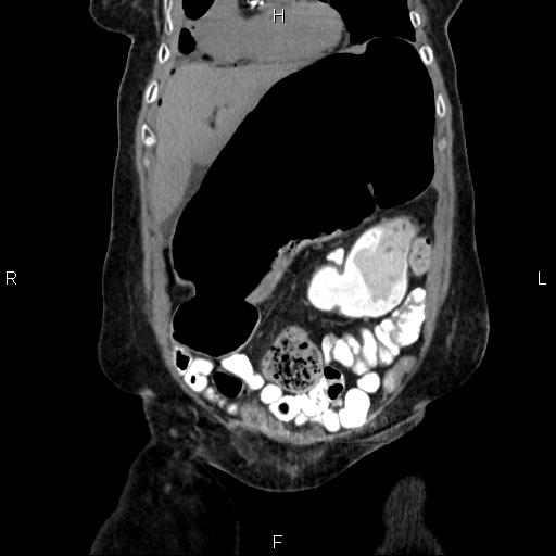
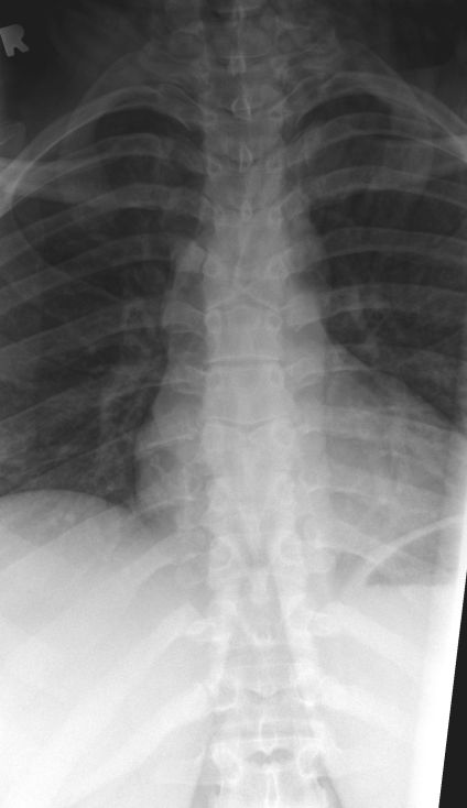
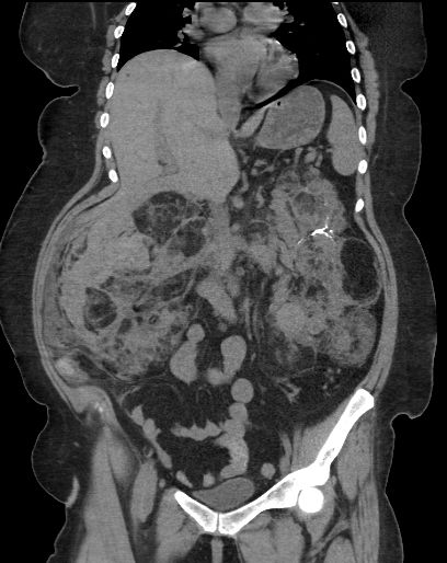
#Wetread Case 19 Answer: Cecal volvulus
= torsion of the cecum around it's mesentery
~10% of intestinal volvuli
30-60yo
often prior abd surgery or pelvic mass
present as prox colon obstruction (pain,n,v, distention)
#FOAMrad #FOAMed #radres #errad #bodyrad

= torsion of the cecum around it's mesentery
~10% of intestinal volvuli
30-60yo
often prior abd surgery or pelvic mass
present as prox colon obstruction (pain,n,v, distention)
#FOAMrad #FOAMed #radres #errad #bodyrad


Cecal volulus:
2 types:
-Axial - twists about axial plane (either way) but remains in RLQ
- Loop type - twists and inverts moving to LUQ
Bascule is a variant where the cecum doesn't twist, just folds up anteriorly (NO torsion!)
From UpToDate (a=axial, b=loop, c=bascule):
2 types:
-Axial - twists about axial plane (either way) but remains in RLQ
- Loop type - twists and inverts moving to LUQ
Bascule is a variant where the cecum doesn't twist, just folds up anteriorly (NO torsion!)
From UpToDate (a=axial, b=loop, c=bascule):

Cecal Volulus:
X-ray: marked dilated colon loop extending from RLQ to LUQ (remember cecum dilation is >9cm)
-haustra usually maintained
-can have SINGLE air-fluid level
CT: exactly what you expect - dilated cecum with "bird beak" at torsion/obstruction
X-ray: marked dilated colon loop extending from RLQ to LUQ (remember cecum dilation is >9cm)
-haustra usually maintained
-can have SINGLE air-fluid level
CT: exactly what you expect - dilated cecum with "bird beak" at torsion/obstruction
Cecal Volvulus:
Treatment:
Surgery vs colonscopic decompression
Look for wall thickening, pneumotosis, free air, arterial cut-offs or venous dilation/obstruction - all concerning signs for ischemia
Often when mesentery twists it pulls in other loops (see sigmoid below)
Treatment:
Surgery vs colonscopic decompression
Look for wall thickening, pneumotosis, free air, arterial cut-offs or venous dilation/obstruction - all concerning signs for ischemia
Often when mesentery twists it pulls in other loops (see sigmoid below)
Cecal vs Sigmoid volvulus
Not always as simple as it sounds.
1) Loop for straight (cecal) vs upside down U-shaped (sigmoid) dilated colon loop
2) Is the descending colon decompressed (cecal) or dilated (sigmoid)?
#FOAMrad #FOAMed #radres #bodyrad


Not always as simple as it sounds.
1) Loop for straight (cecal) vs upside down U-shaped (sigmoid) dilated colon loop
2) Is the descending colon decompressed (cecal) or dilated (sigmoid)?
#FOAMrad #FOAMed #radres #bodyrad


• • •
Missing some Tweet in this thread? You can try to
force a refresh


